
















Copyright: ©Author(s) 2026.
World J Clin Cases. Jun 26, 2026; 14(18): 120279
Published online Jun 26, 2026. doi: 10.12998/wjcc.120279
Published online Jun 26, 2026. doi: 10.12998/wjcc.120279
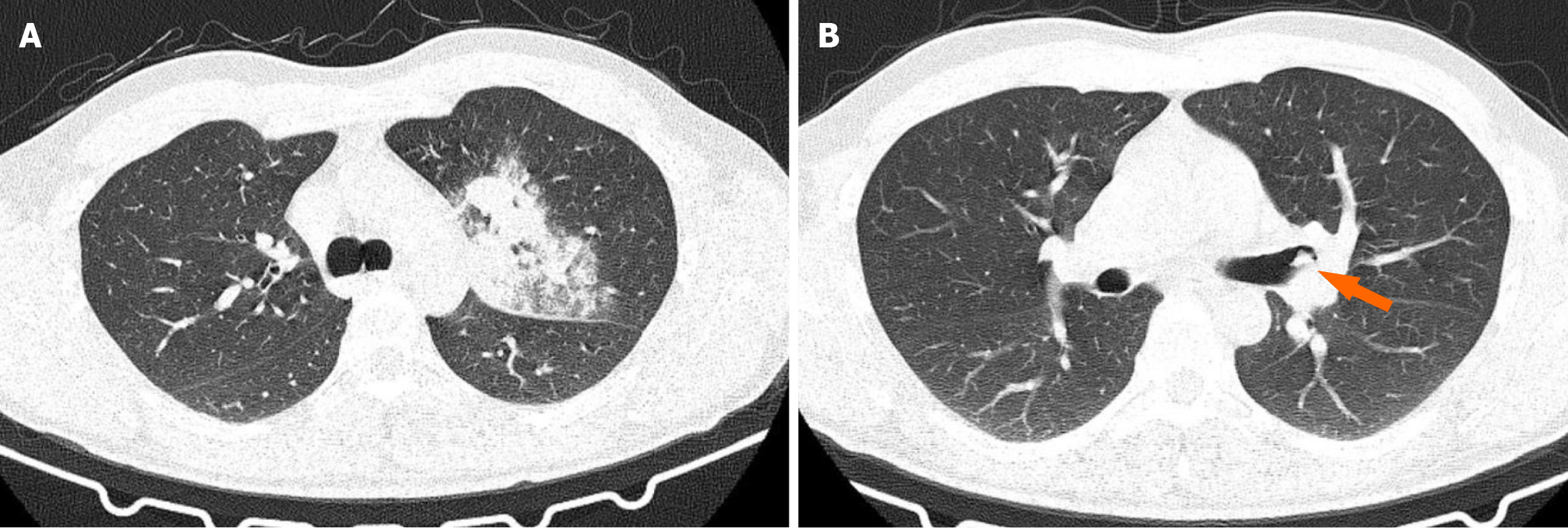
Figure 1 Chest computed tomography result.
A: Left upper lobe consolidation secondary to endobronchial obstruction. Axial chest computed tomography (CT; lung window) showing consolidation with air bronchograms in the left upper lobe, associated with surrounding ground-glass opacities. The imaging findings are consistent with post-obstructive pneumonia related to an underlying endobronchial lesion located between the left main bronchus and the left upper lobar bronchus (not shown in this slice); B: Endobronchial chondroma. Axial chest CT image showing an endobronchial lesion located between the left main bronchus and the left upper lobar bronchus (arrow), corresponding to the obstructing mass identified on bronchoscopy.
- Citation: Dodos K, Kalamara TV, Georgakopoulou VE. Endobronchial chondroma presenting as recurrent pneumonia - diagnostic role of bronchoscopy and surgical management: A case report. World J Clin Cases 2026; 14(18): 120279
- URL: https://www.wjgnet.com/2307-8960/full/v14/i18/120279.htm
- DOI: https://dx.doi.org/10.12998/wjcc.120279