
















Copyright: ©Author(s) 2026.
World J Clin Cases. Jun 6, 2026; 14(16): 120594
Published online Jun 6, 2026. doi: 10.12998/wjcc.v14.i16.120594
Published online Jun 6, 2026. doi: 10.12998/wjcc.v14.i16.120594
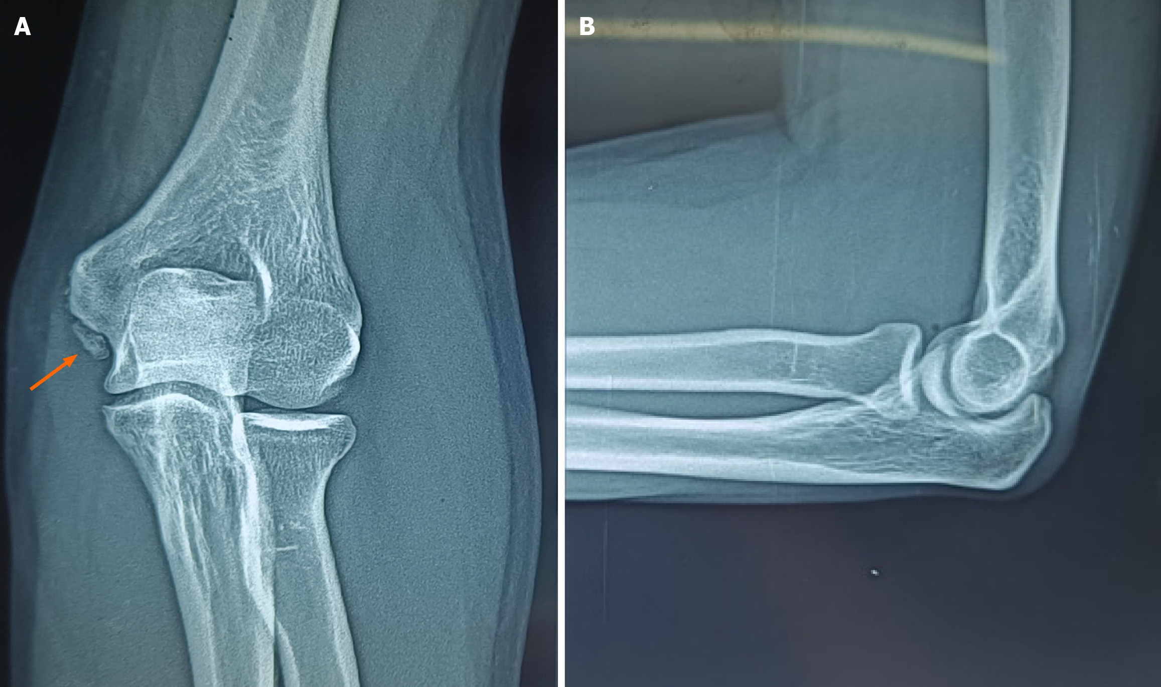
Figure 1 Plain radiograph of the left elbow.
A and B: Anteroposterior and lateral view. The orange arrow indicates an accessory bone located beneath the medial epicondyle, featuring a distinctive, well-defined oval ossicle with smooth margins.
- Citation: Nallakumarasamy A, Sivakumar S, Vetrivel VN, Balaji VP, Stephen N, Jeyaraman N, Jeyaraman M. Os subepicondylare mediale ossicle as a cause for cubital tunnel syndrome: A case report. World J Clin Cases 2026; 14(16): 120594
- URL: https://www.wjgnet.com/2307-8960/full/v14/i16/120594.htm
- DOI: https://dx.doi.org/10.12998/wjcc.v14.i16.120594