
















Copyright: ©Author(s) 2026.
World J Clin Cases. Jun 6, 2026; 14(16): 120049
Published online Jun 6, 2026. doi: 10.12998/wjcc.v14.i16.120049
Published online Jun 6, 2026. doi: 10.12998/wjcc.v14.i16.120049
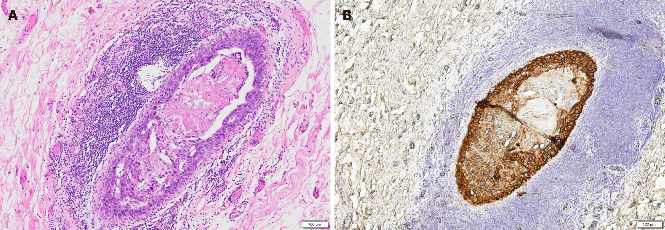
Figure 6 High-grade ductal carcinoma in situ in the partial mastectomy specimen.
A and B: Representative sections are shown which demonstrated monotonous tumor cells with enlarged, hyperchromatic nuclei filling and expanding mammary ducts. Central comedo-type necrosis and peripheral reactive lymphoid aggregates were present, consistent with high-grade ductal carcinoma in situ.
- Citation: Tsai CY, Lo TC, Yu JC, Hong ZJ, Sung YK. Paget’s disease of the nipple with occult ductal carcinoma in situ despite negative conventional imaging: A case report. World J Clin Cases 2026; 14(16): 120049
- URL: https://www.wjgnet.com/2307-8960/full/v14/i16/120049.htm
- DOI: https://dx.doi.org/10.12998/wjcc.v14.i16.120049