
















Copyright: ©Author(s) 2026.
World J Clin Cases. Jun 6, 2026; 14(16): 120049
Published online Jun 6, 2026. doi: 10.12998/wjcc.v14.i16.120049
Published online Jun 6, 2026. doi: 10.12998/wjcc.v14.i16.120049
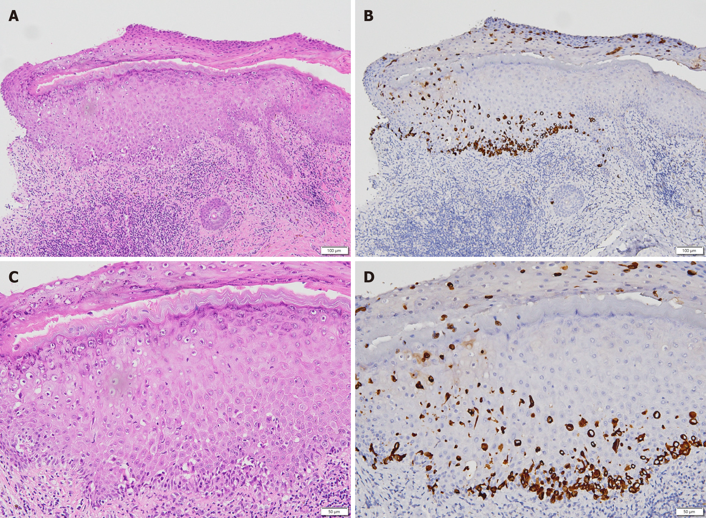
Figure 5 Histopathologic features of Paget’s disease of the nipple.
A: Skin, 40 µm; B: Cytokeratin 7 (CK7) immunostaining, 40 µm; C: Skin, 100 µm; D: CK7 immunostaining, 100 µm. Microscopic examination demonstrated malignant ductal epithelial cells spreading along the basal layer and infiltrating upward into the overlying squamous epithelium. Paget cells were ovoid, with high nuclear-to-cytoplasmic ratios, hyperchromatic nuclei, and pale cytoplasm. Immunohistochemistry showed strong CK7 positivity, supporting the diagnosis.
- Citation: Tsai CY, Lo TC, Yu JC, Hong ZJ, Sung YK. Paget’s disease of the nipple with occult ductal carcinoma in situ despite negative conventional imaging: A case report. World J Clin Cases 2026; 14(16): 120049
- URL: https://www.wjgnet.com/2307-8960/full/v14/i16/120049.htm
- DOI: https://dx.doi.org/10.12998/wjcc.v14.i16.120049