
















Copyright: ©Author(s) 2026.
World J Clin Cases. Jun 6, 2026; 14(16): 120049
Published online Jun 6, 2026. doi: 10.12998/wjcc.v14.i16.120049
Published online Jun 6, 2026. doi: 10.12998/wjcc.v14.i16.120049
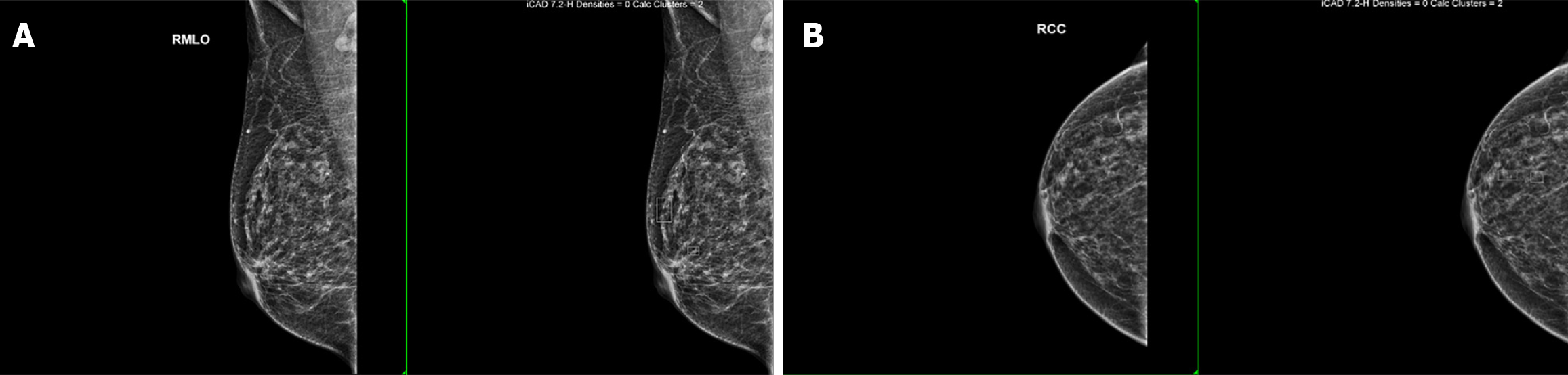
Figure 2 Breast mammography.
A: Right breast mammography. Right craniocaudal and right mediolateral oblique views demonstrated grouped punctate and amorphous calcifications in the upper outer quadrant and retroareolar region of the right breast, without associated mass, architectural distortion, or lym
- Citation: Tsai CY, Lo TC, Yu JC, Hong ZJ, Sung YK. Paget’s disease of the nipple with occult ductal carcinoma in situ despite negative conventional imaging: A case report. World J Clin Cases 2026; 14(16): 120049
- URL: https://www.wjgnet.com/2307-8960/full/v14/i16/120049.htm
- DOI: https://dx.doi.org/10.12998/wjcc.v14.i16.120049