
















Copyright: ©Author(s) 2026.
World J Clin Cases. May 26, 2026; 14(15): 120825
Published online May 26, 2026. doi: 10.12998/wjcc.v14.i15.120825
Published online May 26, 2026. doi: 10.12998/wjcc.v14.i15.120825
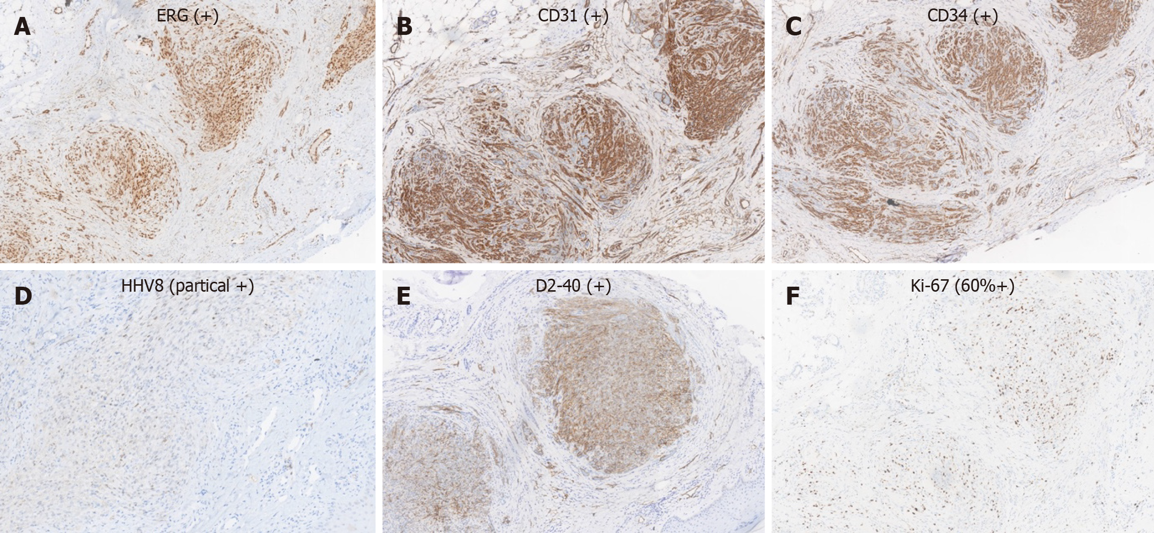
Figure 3 Immunohistochemical staining of Kaposi sarcoma lesions (× 200).
A-F: Representative immunohistochemical staining demonstrating tumor cell markers in Kaposi sarcoma tissue. Tumor cells were positive for ERG (A), CD31 (B), CD34 (C), human herpesvirus-8 (partial positive) (D), D2-40 (E), and Ki67 (approximately 60% positive) (F).
- Citation: Wang SN, Wang JM, Xu M, Wu JJ. Kaposi sarcoma following dupilumab treatment for bullous pemphigoid: A case report and review of literature. World J Clin Cases 2026; 14(15): 120825
- URL: https://www.wjgnet.com/2307-8960/full/v14/i15/120825.htm
- DOI: https://dx.doi.org/10.12998/wjcc.v14.i15.120825