
















Copyright: ©Author(s) 2026.
World J Clin Cases. May 26, 2026; 14(15): 120269
Published online May 26, 2026. doi: 10.12998/wjcc.v14.i15.120269
Published online May 26, 2026. doi: 10.12998/wjcc.v14.i15.120269
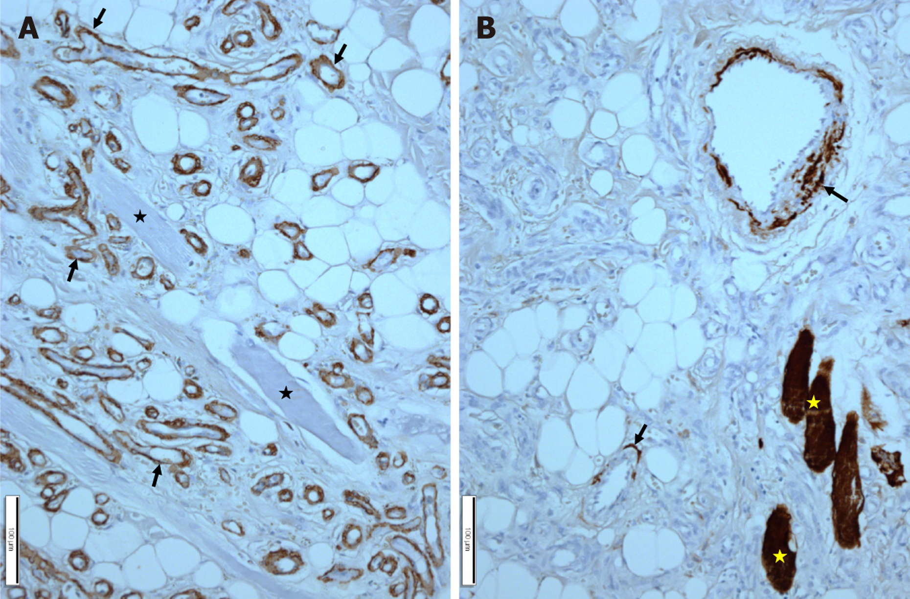
Figure 6 Immunohistochemical staining of the nasal alar base mass.
A: Most of the vascular wall (arrows) shows positive immunoreactivity for the alpha-smooth muscle actin. Entrapped skeletal muscle fibers (asterisks) show negative immunoreactivity; B: Most of the small blood vessels show negative immunoreactivity for desmin, but large sized blood vessels (arrows) and entrapped skeletal muscle fibers (asterisks) show positive immunoreactivity. Scale bars measure 100 μm.
- Citation: Choi JY, Aldosari AM, Lim SC. Angiolipoma of the nasal alar base: A case report. World J Clin Cases 2026; 14(15): 120269
- URL: https://www.wjgnet.com/2307-8960/full/v14/i15/120269.htm
- DOI: https://dx.doi.org/10.12998/wjcc.v14.i15.120269