
















Copyright: ©Author(s) 2026.
World J Clin Cases. May 26, 2026; 14(15): 120269
Published online May 26, 2026. doi: 10.12998/wjcc.v14.i15.120269
Published online May 26, 2026. doi: 10.12998/wjcc.v14.i15.120269
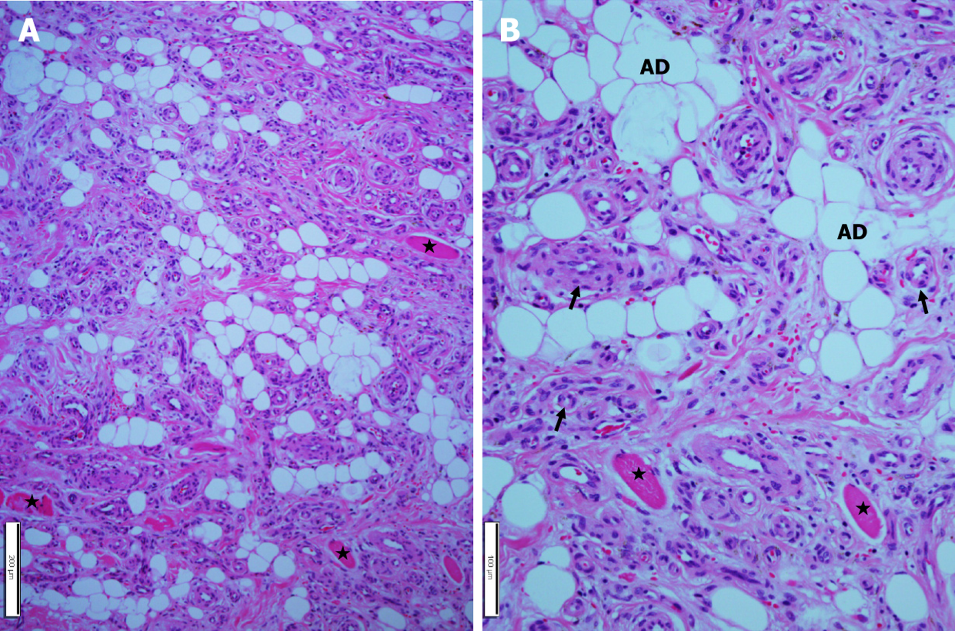
Figure 5 Histopathologic findings of the nasal alar base mass.
A: The tumor comprises a lot of blood vessels, adipocytes, and entrapped skeletal muscle fibers (asterisks); B: Higher magnification shows thin- and thick-walled blood vessels (arrows) and mature adipocytes. No cytological atypica or mitosis. Entrapped skeletal muscle fibers (asterisks) are noted. Hematoxylin and eosin staining. Scale bars measure A: 200 μm; B: 100 μm. AD: Adipocytes.
- Citation: Choi JY, Aldosari AM, Lim SC. Angiolipoma of the nasal alar base: A case report. World J Clin Cases 2026; 14(15): 120269
- URL: https://www.wjgnet.com/2307-8960/full/v14/i15/120269.htm
- DOI: https://dx.doi.org/10.12998/wjcc.v14.i15.120269