
















Copyright: ©Author(s) 2026.
World J Clin Cases. May 26, 2026; 14(15): 120061
Published online May 26, 2026. doi: 10.12998/wjcc.v14.i15.120061
Published online May 26, 2026. doi: 10.12998/wjcc.v14.i15.120061
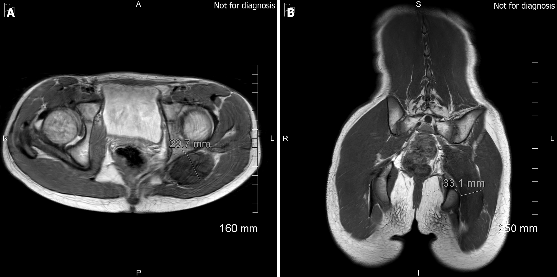
Figure 3 Magnetic resonance imaging of the lumbosacral region from November 2025.
A: Axial image at the level of the ischium showing an anteroposterior sciatic nerve diameter of approximately 3.0 cm; B: Coronal image at the level of the ischial tuberosity showing a transverse diameter of approximately 3.3 cm. Overall appearances were similar to prior studies, with no convincing interval progression in morphology or signal characteristics.
- Citation: Saravanan K, Nagappan PG, Simpson AI. Benign but not inconsequential, paediatric sciatic nerve neuromuscular choristoma and its longitudinal impact: A case report. World J Clin Cases 2026; 14(15): 120061
- URL: https://www.wjgnet.com/2307-8960/full/v14/i15/120061.htm
- DOI: https://dx.doi.org/10.12998/wjcc.v14.i15.120061