
















Copyright: ©Author(s) 2026.
World J Clin Cases. May 26, 2026; 14(15): 120061
Published online May 26, 2026. doi: 10.12998/wjcc.v14.i15.120061
Published online May 26, 2026. doi: 10.12998/wjcc.v14.i15.120061
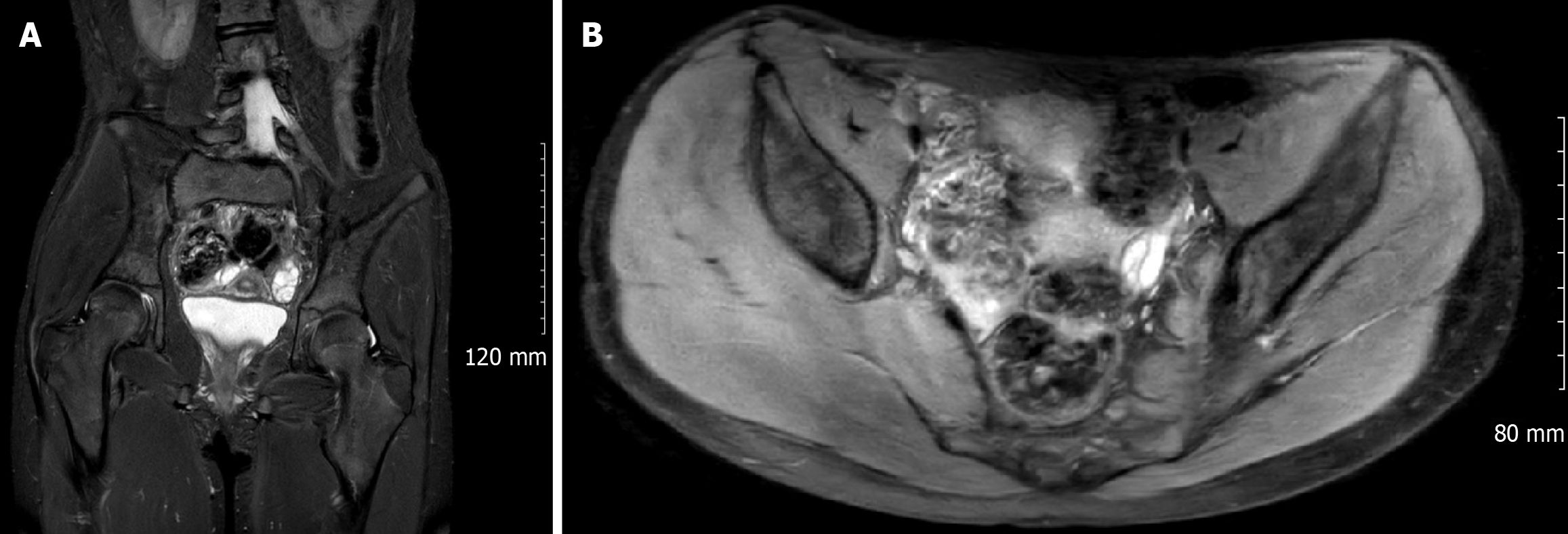
Figure 2 Magnetic resonance imaging of the pelvis (August 2024) focusing on the lumbosacral exit foramina.
The images highlight enlargement of the neural foramina and a thickened left L5 nerve root (seen in cross-section) caused by the choristoma. There is smooth expansion of the nerve root sleeve (outlined) and no destructive bony changes, consistent with a chronic benign process that leads to widening of the nerve root canals without invasive or infiltrative features. A: Axial view; B: Coronal view.
- Citation: Saravanan K, Nagappan PG, Simpson AI. Benign but not inconsequential, paediatric sciatic nerve neuromuscular choristoma and its longitudinal impact: A case report. World J Clin Cases 2026; 14(15): 120061
- URL: https://www.wjgnet.com/2307-8960/full/v14/i15/120061.htm
- DOI: https://dx.doi.org/10.12998/wjcc.v14.i15.120061