
















Copyright: ©Author(s) 2026.
World J Clin Cases. May 26, 2026; 14(15): 120061
Published online May 26, 2026. doi: 10.12998/wjcc.v14.i15.120061
Published online May 26, 2026. doi: 10.12998/wjcc.v14.i15.120061
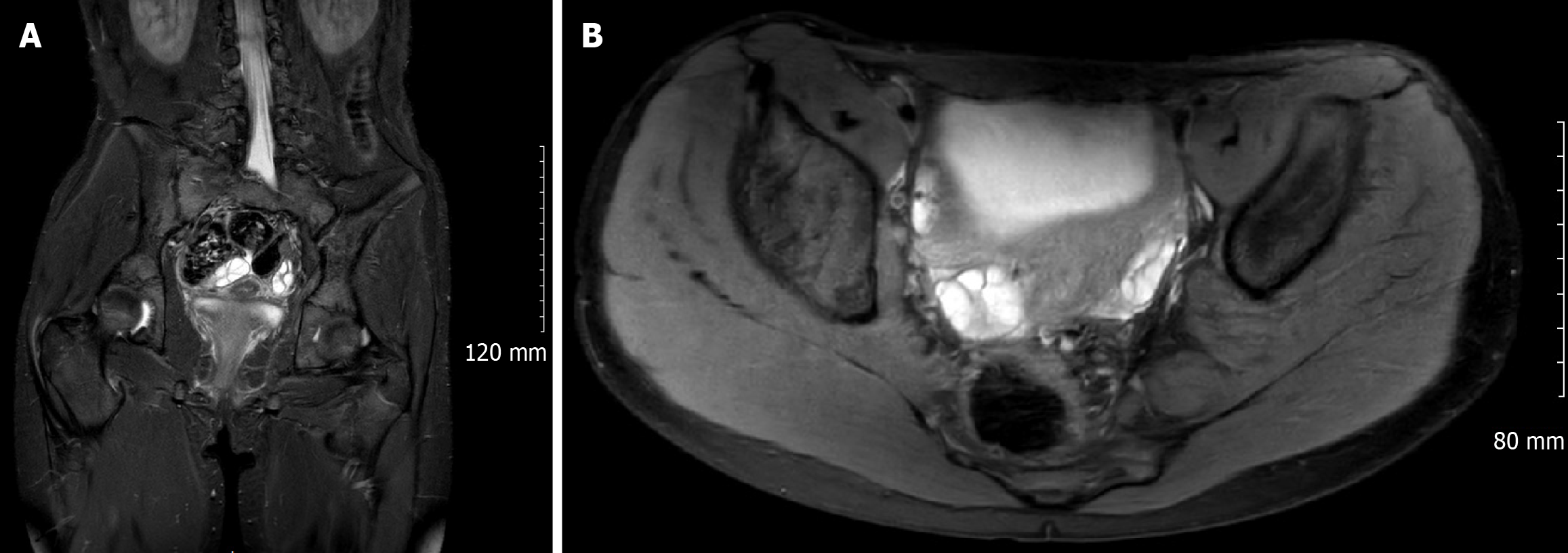
Figure 1 T2-weighted magnetic resonance imaging of the lumbosacral region (August 2024) demonstrating nerve root enlargement due to neuromuscular choristoma.
A: Expansion of the left L4–S2 nerve roots through the pelvis, indicated by arrowheads; B: Axial magnetic resonance imaging view at the level of the sciatic nerve, illustrating markedly hypertrophic left sciatic nerve fascicles (encircled) on the left side. These images confirm the findings of sciatic nerve neuromuscular choristoma without evidence of a discrete mass or malignant change.
- Citation: Saravanan K, Nagappan PG, Simpson AI. Benign but not inconsequential, paediatric sciatic nerve neuromuscular choristoma and its longitudinal impact: A case report. World J Clin Cases 2026; 14(15): 120061
- URL: https://www.wjgnet.com/2307-8960/full/v14/i15/120061.htm
- DOI: https://dx.doi.org/10.12998/wjcc.v14.i15.120061