
















Copyright: ©Author(s) 2026.
World J Clin Cases. May 16, 2026; 14(14): 119746
Published online May 16, 2026. doi: 10.12998/wjcc.v14.i14.119746
Published online May 16, 2026. doi: 10.12998/wjcc.v14.i14.119746
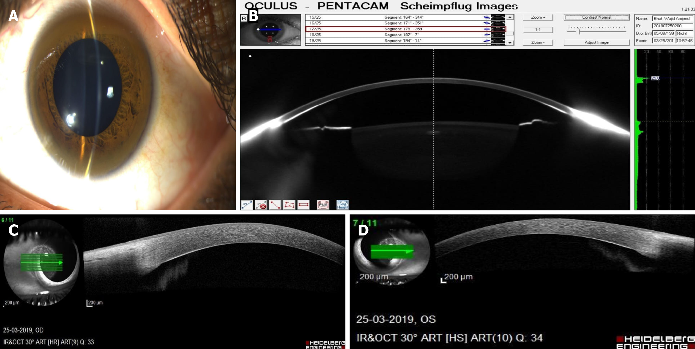
Figure 2 Imaging modalities used for detection of Kayser-Fleischer ring in Wilson’s disease.
A: Slit-lamp photograph demonstrating complete circumferential Kayser-Fleischer (KF) ring pigmentation at the limbus; B: Scheimpflug imaging showing increased peripheral corneal density corresponding to copper deposition; C: Anterior segment optical coherence tomography (AS-OCT) demonstrating a peripheral hyper-reflective band at the level of Descemet’s membrane consistent with KF ring in right eye; D: AS-OCT image illustrating circumferential hyper-reflective deposition along Descemet’s membrane, corresponding to corneal copper accumulation in left eye. OCT: Optical coherence tomography; ART: Ambrosio’s relational thickness; OS: Oculus sinister; OD: Oculus dexter; IR: Infrared; HS: High speed.
- Citation: Kaur M, Walia S, Morya AK, Bang PH, Aggarwal S, Gupta PC, Nagare R, Udenia H, Khullar S, Morya R. Pathophysiology of ocular manifestations in Wilson’s disease and its management. World J Clin Cases 2026; 14(14): 119746
- URL: https://www.wjgnet.com/2307-8960/full/v14/i14/119746.htm
- DOI: https://dx.doi.org/10.12998/wjcc.v14.i14.119746