
















Copyright: ©Author(s) 2026.
World J Clin Cases. May 16, 2026; 14(14): 119746
Published online May 16, 2026. doi: 10.12998/wjcc.v14.i14.119746
Published online May 16, 2026. doi: 10.12998/wjcc.v14.i14.119746
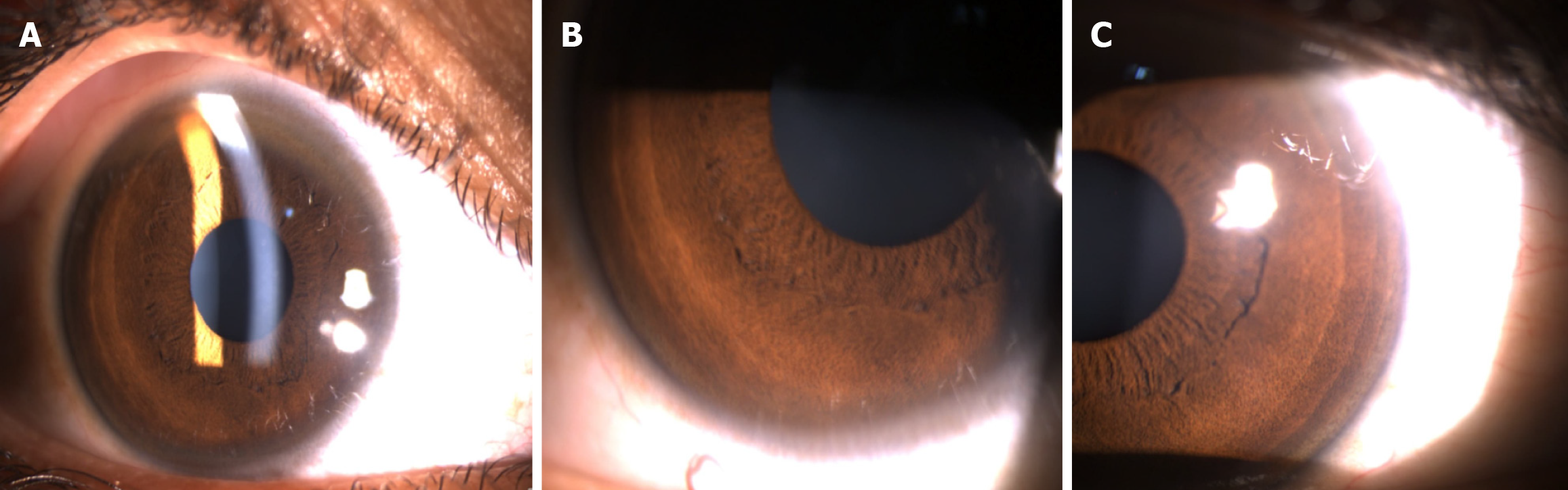
Figure 1 Kayser-Fleischer ring progression in Wilson’s disease.
A: Early Kayser-Fleischer (KF) ring appearing as a superior peripheral corneal arc on slit-lamp examination; B: Inferior corneal involvement due to progressive copper deposition; C: Complete circumferential KF ring at the corneal periphery.
- Citation: Kaur M, Walia S, Morya AK, Bang PH, Aggarwal S, Gupta PC, Nagare R, Udenia H, Khullar S, Morya R. Pathophysiology of ocular manifestations in Wilson’s disease and its management. World J Clin Cases 2026; 14(14): 119746
- URL: https://www.wjgnet.com/2307-8960/full/v14/i14/119746.htm
- DOI: https://dx.doi.org/10.12998/wjcc.v14.i14.119746