
















Copyright: ©Author(s) 2026.
World J Clin Cases. May 6, 2026; 14(13): 117852
Published online May 6, 2026. doi: 10.12998/wjcc.v14.i13.117852
Published online May 6, 2026. doi: 10.12998/wjcc.v14.i13.117852
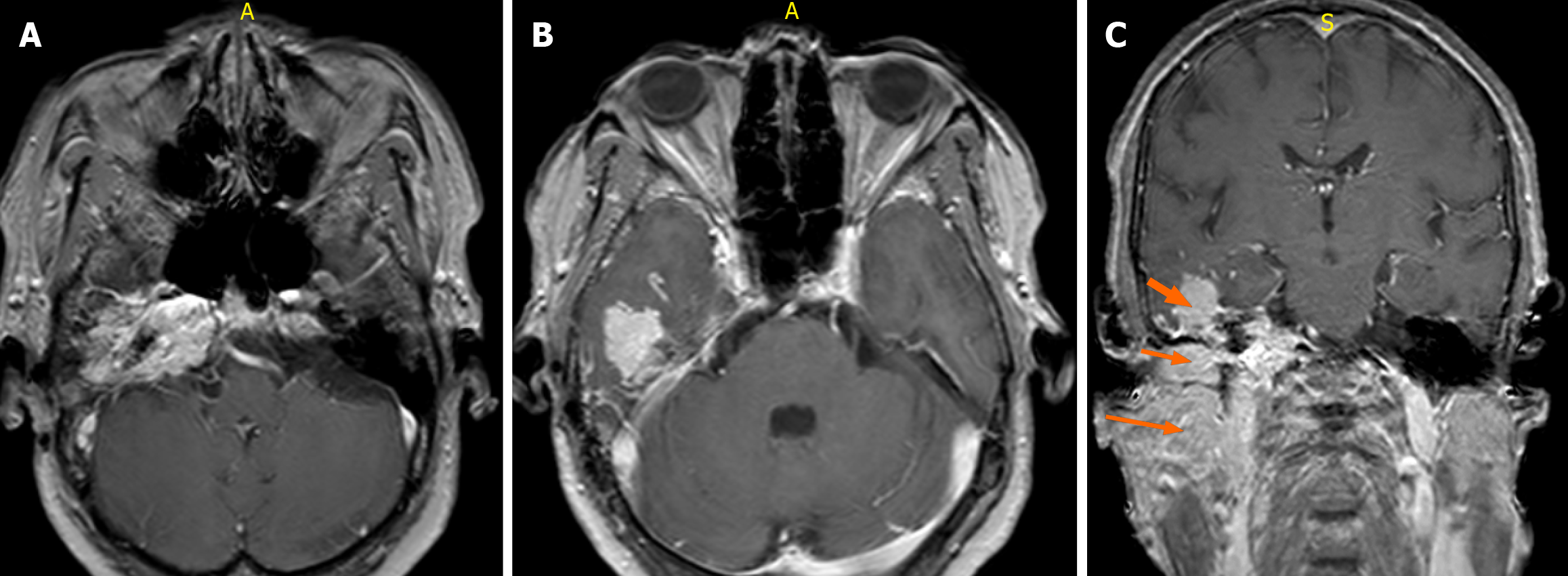
Figure 5 Contrast-enhanced magnetic resonance imaging features of the right jugular foramen mass.
A and B: Axial gadolinium-enhanced T1-weighted fast field echo (FFE) magnetic resonance imaging (repetition time 500-700 milliseconds, echo time 10-20 milliseconds, flip angle 20°-30°, slice thickness 3 mm) following intravenous administration of gadolinium-based contrast (0.1 mmol/kg) demonstrates avid enhancement of the lesion involving the right jugular foramen, middle ear cavity, and ipsilateral temporal lobe; C: Coronal T1-weighted FFE post-contrast image shows an intensely enhancing mass in the right jugular fossa with superolateral extension into the ipsilateral inferior temporal lobe (short thick arrow), inferior extension into the ipsilateral middle ear cavity (short thin arrow), and further spread into the masticator space (long arrow).
- Citation: Patel H, Okuna Y, Ujima W. Atypical jugulotympanic paraganglioma presentation, diagnostic challenges, and role of imaging: A case report. World J Clin Cases 2026; 14(13): 117852
- URL: https://www.wjgnet.com/2307-8960/full/v14/i13/117852.htm
- DOI: https://dx.doi.org/10.12998/wjcc.v14.i13.117852