
















Copyright: ©Author(s) 2026.
World J Clin Cases. May 6, 2026; 14(13): 117852
Published online May 6, 2026. doi: 10.12998/wjcc.v14.i13.117852
Published online May 6, 2026. doi: 10.12998/wjcc.v14.i13.117852
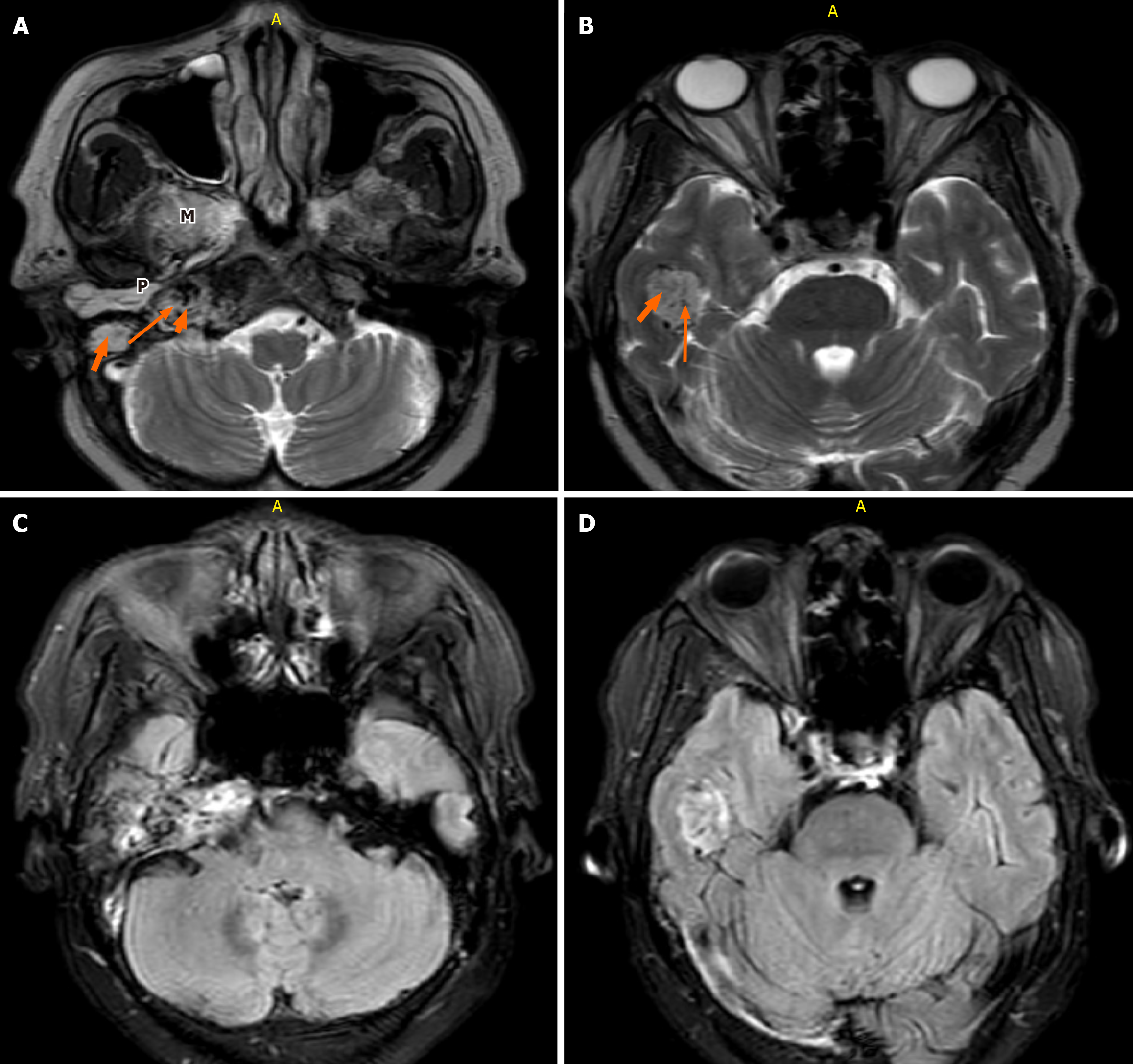
Figure 4 T2-weighted magnetic resonance imaging.
A and B: Axial T2-weighted magnetic resonance imaging of the brain (fast spin echo sequence; repetition time 4500-6000 milliseconds, echo time 90-110 milliseconds, 3-mm slice thickness) demonstrates a slightly hyperintense, heterogeneous mass in the region of the right jugular foramen (long arrow). A characteristic “salt and pepper” appearance is noted. The lesion extends inferiorly into the ipsilateral middle ear cavity (short thick arrow) and right masticator space (M), with superior extension into the ipsilateral temporal lobe (arrows). Hyperintense fluid signal within the right mastoid air cells indicates retained secretions (P); C and D: Axial fluid-attenuated inversion recovery magnetic resonance imaging shows a heterogeneous hypo- to hyperintense lesion involving the right jugular foramen and extending into the ipsilateral temporal lobe.
- Citation: Patel H, Okuna Y, Ujima W. Atypical jugulotympanic paraganglioma presentation, diagnostic challenges, and role of imaging: A case report. World J Clin Cases 2026; 14(13): 117852
- URL: https://www.wjgnet.com/2307-8960/full/v14/i13/117852.htm
- DOI: https://dx.doi.org/10.12998/wjcc.v14.i13.117852