
















Copyright: ©Author(s) 2026.
World J Clin Cases. May 6, 2026; 14(13): 117852
Published online May 6, 2026. doi: 10.12998/wjcc.v14.i13.117852
Published online May 6, 2026. doi: 10.12998/wjcc.v14.i13.117852
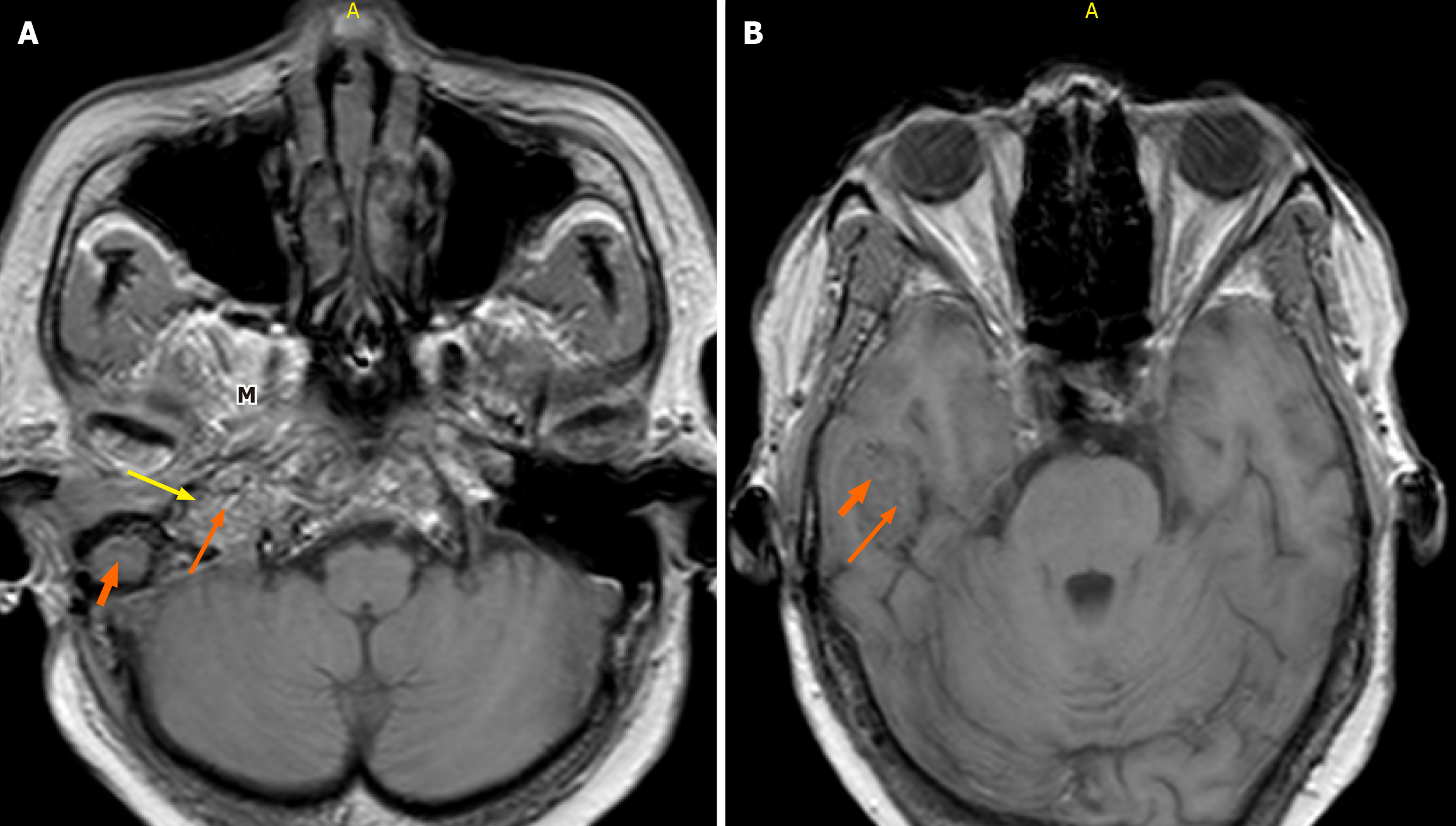
Figure 3 Axial non-contrast-enhanced T1-weighted magnetic resonance imaging of the brain (fast field echo sequence; repetition time 600 milliseconds, echo time 15 milliseconds, flip angle 25°, 3-mm slice thickness).
A and B: Of the brain demonstrates a poorly defined heterogeneous mass of slight hyperintensity and hypointensity centered at the right jugular fossa. A: Characteristic “salt” and “pepper” appearance is observed with punctate regions of hyperintensity representing the “salt” and small flow voids representing the “pepper” (long orange arrow and yellow arrow, respectively). Middle ear extension (thick short arrow) and masticator space (M) are seen; B: The right temporal lobe extension is demonstrated.
- Citation: Patel H, Okuna Y, Ujima W. Atypical jugulotympanic paraganglioma presentation, diagnostic challenges, and role of imaging: A case report. World J Clin Cases 2026; 14(13): 117852
- URL: https://www.wjgnet.com/2307-8960/full/v14/i13/117852.htm
- DOI: https://dx.doi.org/10.12998/wjcc.v14.i13.117852