
















Copyright: ©Author(s) 2026.
World J Clin Cases. May 6, 2026; 14(13): 117852
Published online May 6, 2026. doi: 10.12998/wjcc.v14.i13.117852
Published online May 6, 2026. doi: 10.12998/wjcc.v14.i13.117852
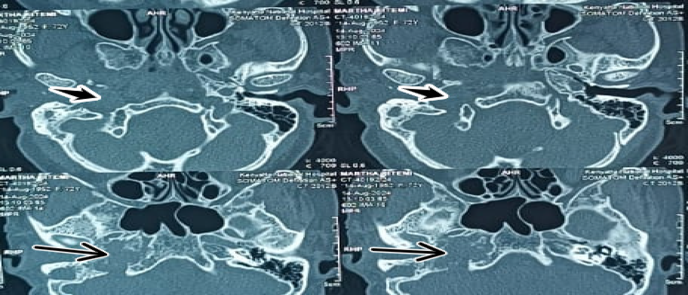
Figure 2 High-resolution non-contrast-enhanced computed tomography of the temporal bone (0.
6-mm slice thickness, bone algorithm reconstruction) showing evidence of prior right mastoidectomy. Permeative bony lesions of the right temporal bone, especially of the temporal apex, right clival region, and right ossicular chain erosion are seen (long thin arrows). The mass is epicentered in the right jugular fossa (short thick arrows).
- Citation: Patel H, Okuna Y, Ujima W. Atypical jugulotympanic paraganglioma presentation, diagnostic challenges, and role of imaging: A case report. World J Clin Cases 2026; 14(13): 117852
- URL: https://www.wjgnet.com/2307-8960/full/v14/i13/117852.htm
- DOI: https://dx.doi.org/10.12998/wjcc.v14.i13.117852