
















Copyright: ©Author(s) 2026.
World J Clin Cases. May 6, 2026; 14(13): 117403
Published online May 6, 2026. doi: 10.12998/wjcc.v14.i13.117403
Published online May 6, 2026. doi: 10.12998/wjcc.v14.i13.117403
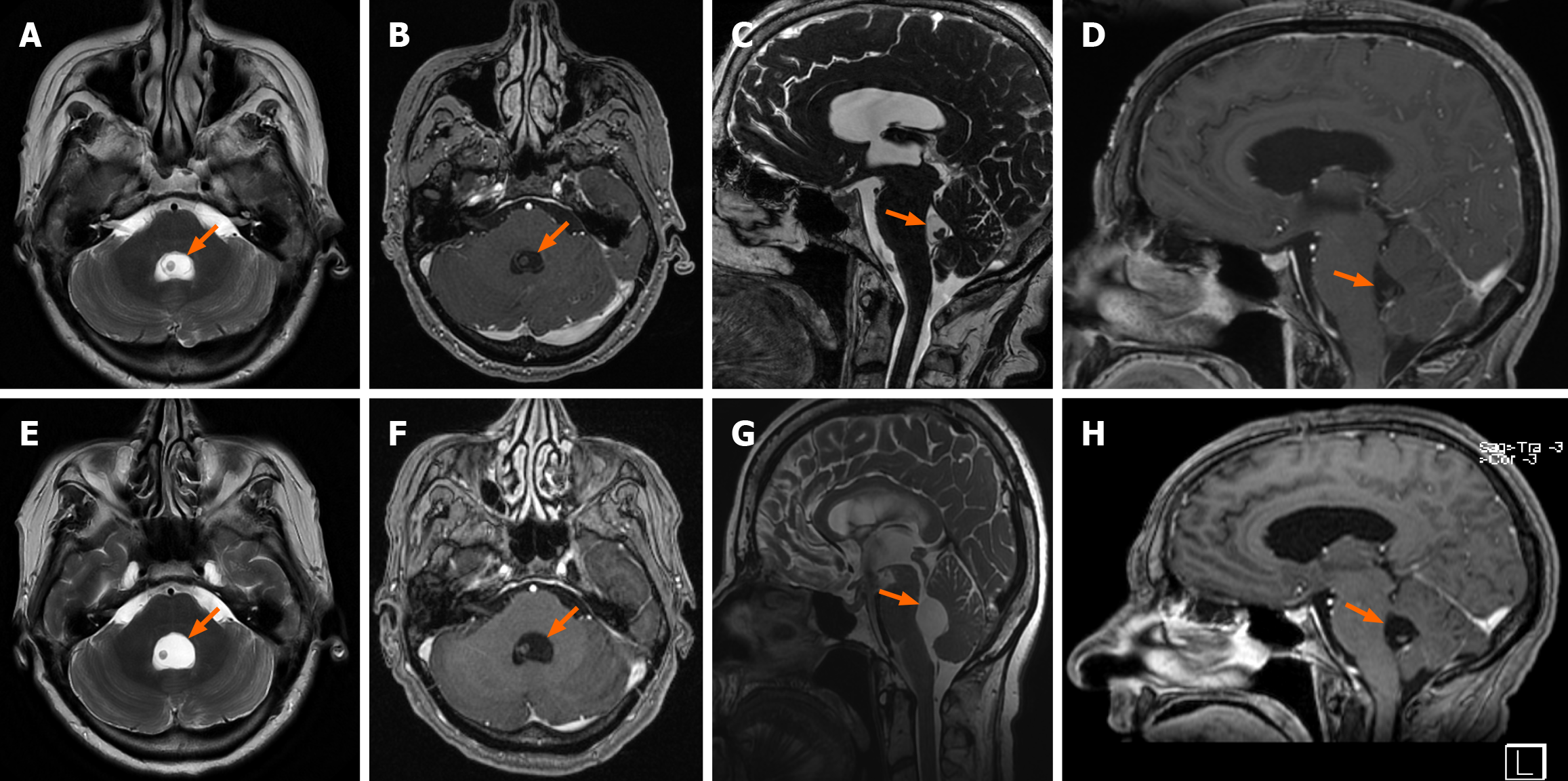
Figure 1 Preoperative magnetic resonance imaging.
A-D: The initial magnetic resonance imaging (MRI) revealed a cystic lesion within the fourth ventricle, characterized by an eccentrically positioned scolex and minimal contrast enhancement. The lesion is indicated by an orange arrow; E-H: A subsequent MRI, performed 5 weeks later, demonstrated a marked increase in the cyst size (A and E: Axial T2-weighted sequences; C and G: Sagittal T2-weighted sequences; B and F: Axial post-contrast sequences; D and H: Sagittal post-contrast sequences).
- Citation: Creatura D, El Choueiri J, Anselmi L, Di Cosmo L, Bono BC, Rampini AD, Lanterna LAA. Fourth ventricle neurocysticercosis with hydrocephalus: A case report and literature review. World J Clin Cases 2026; 14(13): 117403
- URL: https://www.wjgnet.com/2307-8960/full/v14/i13/117403.htm
- DOI: https://dx.doi.org/10.12998/wjcc.v14.i13.117403