
















Copyright: ©Author(s) 2026.
World J Clin Cases. Apr 26, 2026; 14(12): 120145
Published online Apr 26, 2026. doi: 10.12998/wjcc.v14.i12.120145
Published online Apr 26, 2026. doi: 10.12998/wjcc.v14.i12.120145
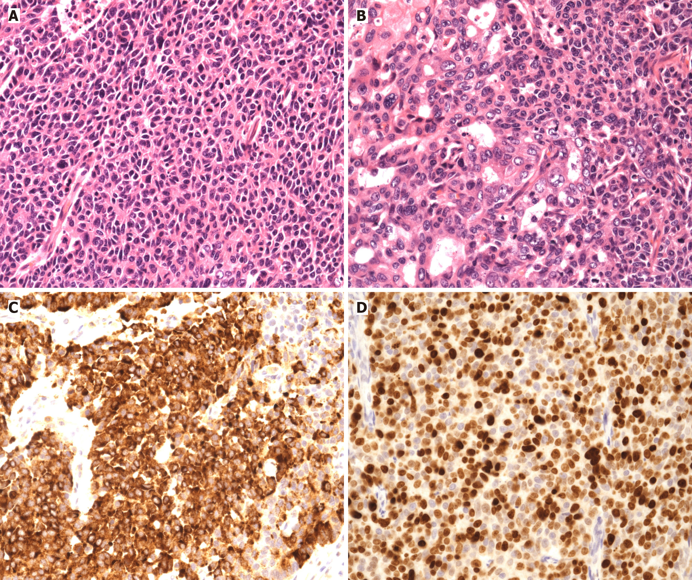
Figure 3 Histopathological findings of large-cell neuroendocrine carcinoma of the ampulla of Vater (× 200).
A: Neoplastic cells with a high nuclear-to-cytoplasmic ratio arranged in solid sheets; B: Focal tubular formation within the tumour, indicating an adenocarcinoma component; C: Diffuse immunohistochemical positivity for chromogranin; D: High Ki-67 proliferation index consistent with a high-grade neuroendocrine carcinoma.
- Citation: Florou E, Velayutham A, Zen Y, Waters J, Srinivasan P. Size-biology paradox in ampullary tumours - large-cell neuroendocrine carcinoma of the ampulla of Vater: A case report. World J Clin Cases 2026; 14(12): 120145
- URL: https://www.wjgnet.com/2307-8960/full/v14/i12/120145.htm
- DOI: https://dx.doi.org/10.12998/wjcc.v14.i12.120145