
















Copyright: ©Author(s) 2026.
World J Clin Cases. Apr 26, 2026; 14(12): 120145
Published online Apr 26, 2026. doi: 10.12998/wjcc.v14.i12.120145
Published online Apr 26, 2026. doi: 10.12998/wjcc.v14.i12.120145
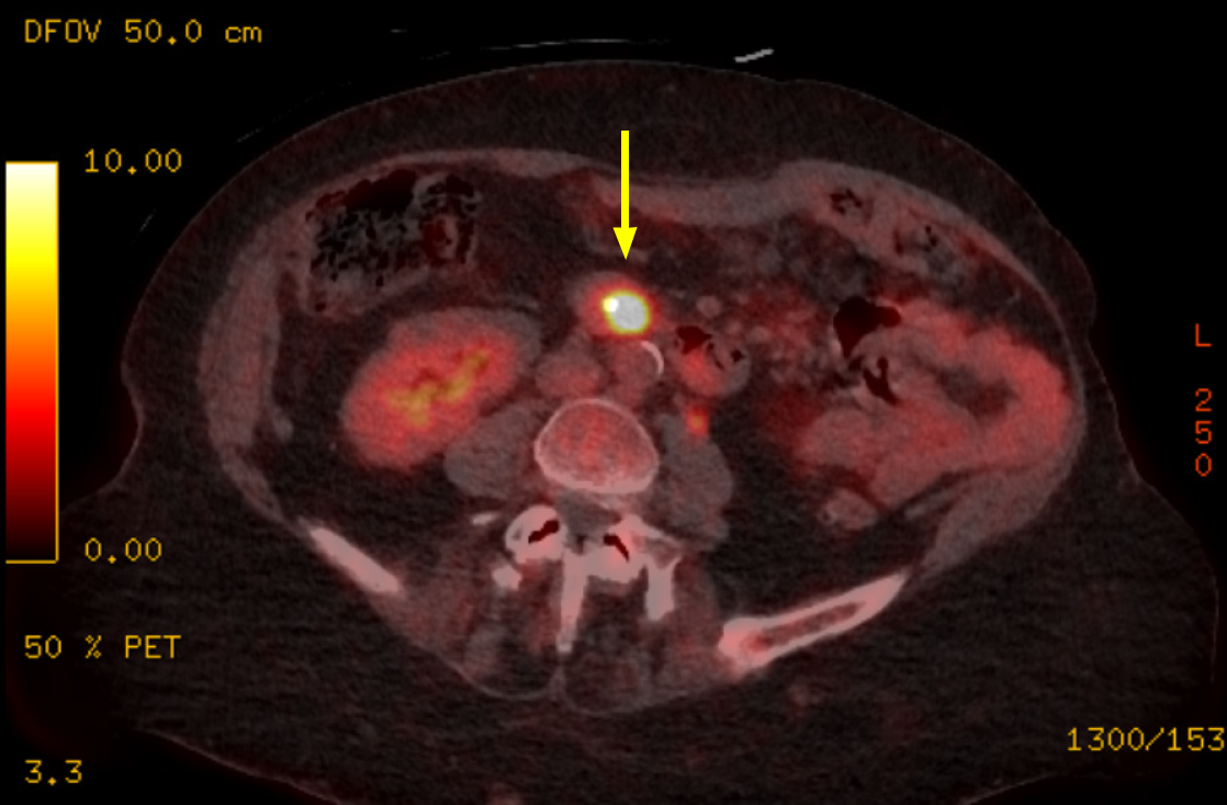
Figure 2 Positron emission tomography/computed tomography.
Axial imaging demonstrates focal fluorodeoxyglucose uptake at the ampulla of Vater (yellow arrow), consistent with a metabolically active ampullary lesion.
- Citation: Florou E, Velayutham A, Zen Y, Waters J, Srinivasan P. Size-biology paradox in ampullary tumours - large-cell neuroendocrine carcinoma of the ampulla of Vater: A case report. World J Clin Cases 2026; 14(12): 120145
- URL: https://www.wjgnet.com/2307-8960/full/v14/i12/120145.htm
- DOI: https://dx.doi.org/10.12998/wjcc.v14.i12.120145