
















Copyright: ©Author(s) 2026.
World J Clin Cases. Apr 26, 2026; 14(12): 118091
Published online Apr 26, 2026. doi: 10.12998/wjcc.v14.i12.118091
Published online Apr 26, 2026. doi: 10.12998/wjcc.v14.i12.118091
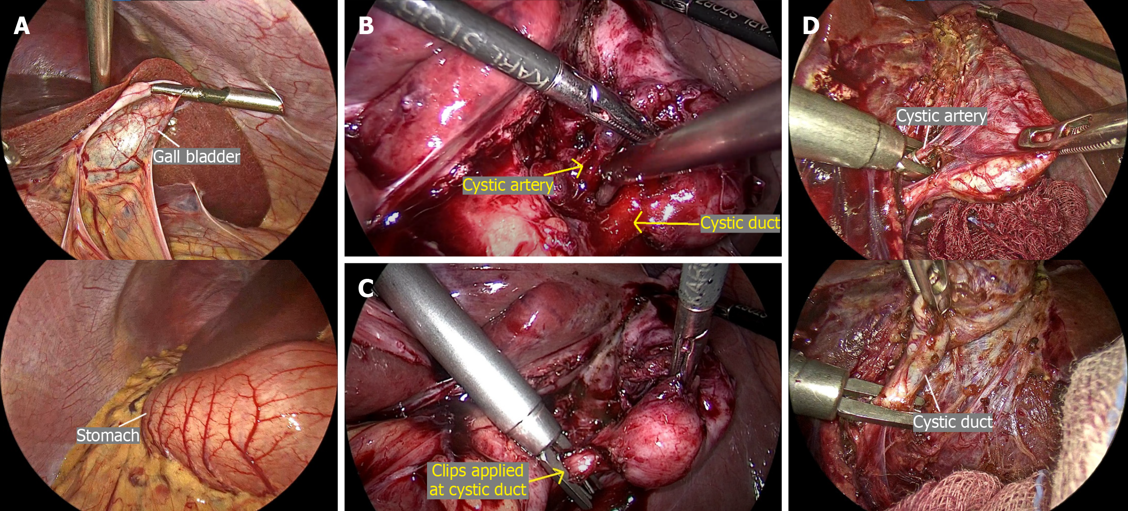
Figure 6 Intraoperative findings during laparoscopic cholecystectomy.
A: Initial laparoscopic assessment demonstrating the liver and gallbladder (white arrow) located in the left hypochondrium, and stomach in the right hypochondrium (white arrow), consistent with situs inversus totalis; B: Dissection of Calot’s triangle. Two tubular structures, corresponding to the cystic duct and cystic artery, are visualized entering the gallbladder (yellow arrow: Cystic artery and duct); C: Control of cystic structures (yellow arrow: Cystic artery and duct). Application of surgical clips to the cystic duct prior to division; D: Critical view of safety. The cystic duct and cystic artery (white arrows) are clearly identified entering the gallbladder, with the lower third of the gallbladder dissected free from the liver bed and complete exposure of Calot’s triangle, following the clinical view of the safety rule.
- Citation: Bhati G, Capolupo GT, Bhati K, Gupta A, Bansal R, Sai Srinija P, Caricato M, Carannante F. Laparoscopic cholecystectomy in patients with situs inversus totalis: Two case reports and review of literature. World J Clin Cases 2026; 14(12): 118091
- URL: https://www.wjgnet.com/2307-8960/full/v14/i12/118091.htm
- DOI: https://dx.doi.org/10.12998/wjcc.v14.i12.118091