















Copyright: ©Author(s) 2026.
World J Clin Cases. Apr 6, 2026; 14(10): 118630
Published online Apr 6, 2026. doi: 10.12998/wjcc.v14.i10.118630
Published online Apr 6, 2026. doi: 10.12998/wjcc.v14.i10.118630
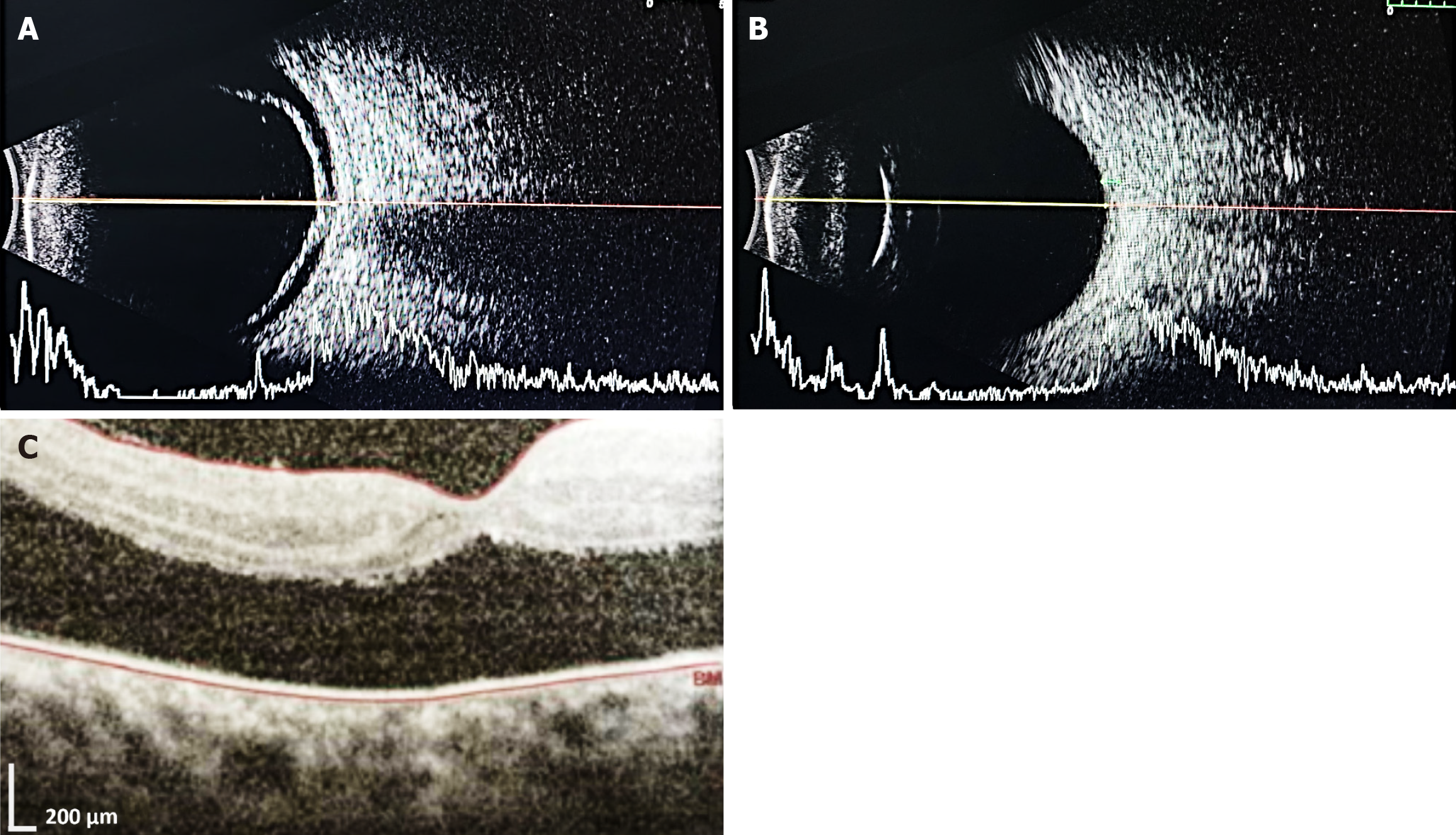
Figure 4 Ocular investigations in case 2 ultrasonography B-scan.
A: Right eye revealing shallow retinal detachment; B: Left eye showing intraocular lens echoes, anechoic vitreous cavity, intact retina-choroid-sclera complex and normal optic nerve head; C: Optical Coherence Tomography of the right eye macula showing a large neurosensory retinal detachment.
- Citation: Raj A, Nishant P, Kumar R, Sharma A, Singh P, Patra PK, Sinha S. Blau syndrome at a tertiary care center of Eastern India: Two case reports and review of literature. World J Clin Cases 2026; 14(10): 118630
- URL: https://www.wjgnet.com/2307-8960/full/v14/i10/118630.htm
- DOI: https://dx.doi.org/10.12998/wjcc.v14.i10.118630