
















©The Author(s) 2026.
World J Clin Cases. Jan 6, 2026; 14(1): 115845
Published online Jan 6, 2026. doi: 10.12998/wjcc.v14.i1.115845
Published online Jan 6, 2026. doi: 10.12998/wjcc.v14.i1.115845
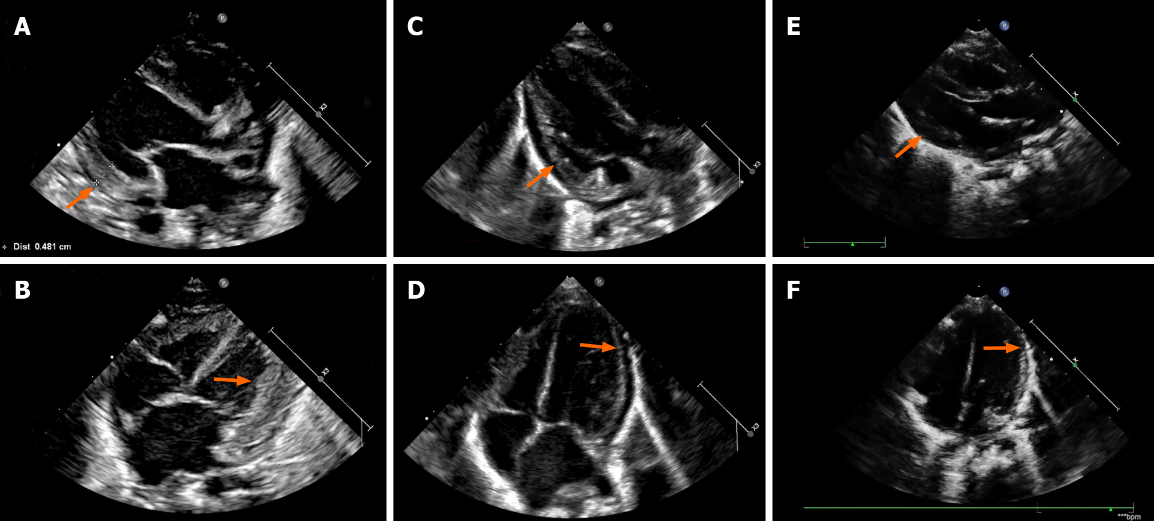
Figure 2 Cardiac color doppler ultrasound changes during levothyroxine treatment.
A and B: Cardiac ultrasound (pre-levothyroxine and 2 days post-levothyroxine) revealed left ventricular hypertrophy and left heart enlargement (orange arrows); C and D: Ultrasound (29 days post-levothyroxine) showed improvement in left heart size and left ventricular hypertrophy (orange arrows); E and F: Ultrasound (92 days post-levothyroxine) showed normalized cardiac structure (orange arrows).
- Citation: Chen HJ, Li J, Xu XM, Zhang B, Cheng BC, Shi J. Preterm heart failure and refractory lactic acidosis caused by congenital hypothyroidism: A case report and review of literature. World J Clin Cases 2026; 14(1): 115845
- URL: https://www.wjgnet.com/2307-8960/full/v14/i1/115845.htm
- DOI: https://dx.doi.org/10.12998/wjcc.v14.i1.115845