
















©The Author(s) 2025.
World J Clin Cases. Dec 26, 2025; 13(36): 113778
Published online Dec 26, 2025. doi: 10.12998/wjcc.v13.i36.113778
Published online Dec 26, 2025. doi: 10.12998/wjcc.v13.i36.113778
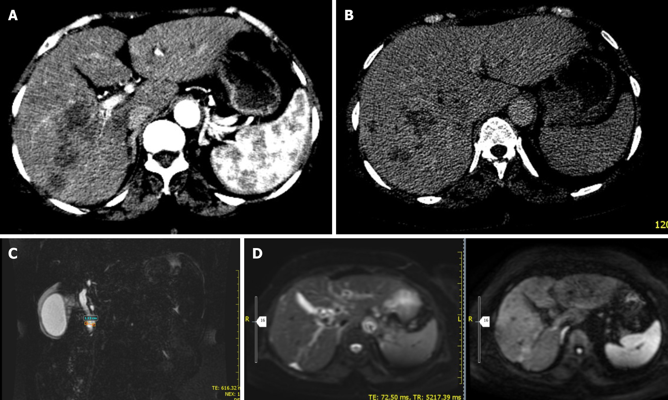
Figure 5 Abdominal computed tomography, magnetic resonance imaging, and magnetic resonance cholangiopancreatography images.
A and B: Shows arterial phase axial abdominal computed tomography (CT) can showing ill defined, confluent hypodense right lobe liver lesions (A). Delayed phase abdominal axial CT scan showing progressive central enhancement of the right lobe hypodense lesion (B); C: Showing dilated complete blood count and edematous gallbladder wall; D: Abdominal magnetic resonance imaging diffusion weighted imaging and apparent diffusion coefficient image showing focal right lobe capsular retraction with adjacent mildly restricting right lobe liver lesion.
- Citation: Mulate ST, Gesese BD, Nur AM, Mengistu HB, Annose RT, Berga AE, Ulfata AL. Hepatic fascioliasis: Emphasizing diagnostic difficulty and the need for high index of suspicion: Four case reports. World J Clin Cases 2025; 13(36): 113778
- URL: https://www.wjgnet.com/2307-8960/full/v13/i36/113778.htm
- DOI: https://dx.doi.org/10.12998/wjcc.v13.i36.113778