
















©The Author(s) 2025.
World J Clin Cases. Dec 26, 2025; 13(36): 113778
Published online Dec 26, 2025. doi: 10.12998/wjcc.v13.i36.113778
Published online Dec 26, 2025. doi: 10.12998/wjcc.v13.i36.113778
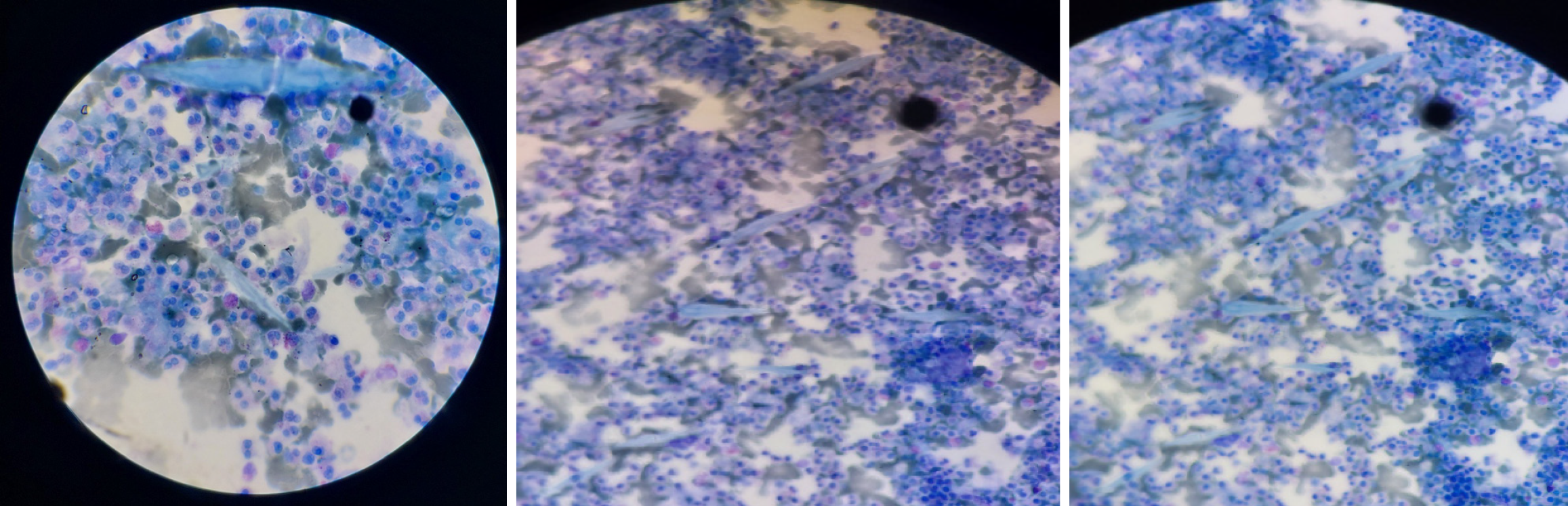
Figure 2 Histopathology of eosinophilic liver abscess.
Photomicrographs demonstrate a necrotic background with sheets of mixed inflammatory cells and scattered Charcot-Leyden crystals.
- Citation: Mulate ST, Gesese BD, Nur AM, Mengistu HB, Annose RT, Berga AE, Ulfata AL. Hepatic fascioliasis: Emphasizing diagnostic difficulty and the need for high index of suspicion: Four case reports. World J Clin Cases 2025; 13(36): 113778
- URL: https://www.wjgnet.com/2307-8960/full/v13/i36/113778.htm
- DOI: https://dx.doi.org/10.12998/wjcc.v13.i36.113778