
















©The Author(s) 2025.
World J Clin Cases. Dec 26, 2025; 13(36): 111835
Published online Dec 26, 2025. doi: 10.12998/wjcc.v13.i36.111835
Published online Dec 26, 2025. doi: 10.12998/wjcc.v13.i36.111835
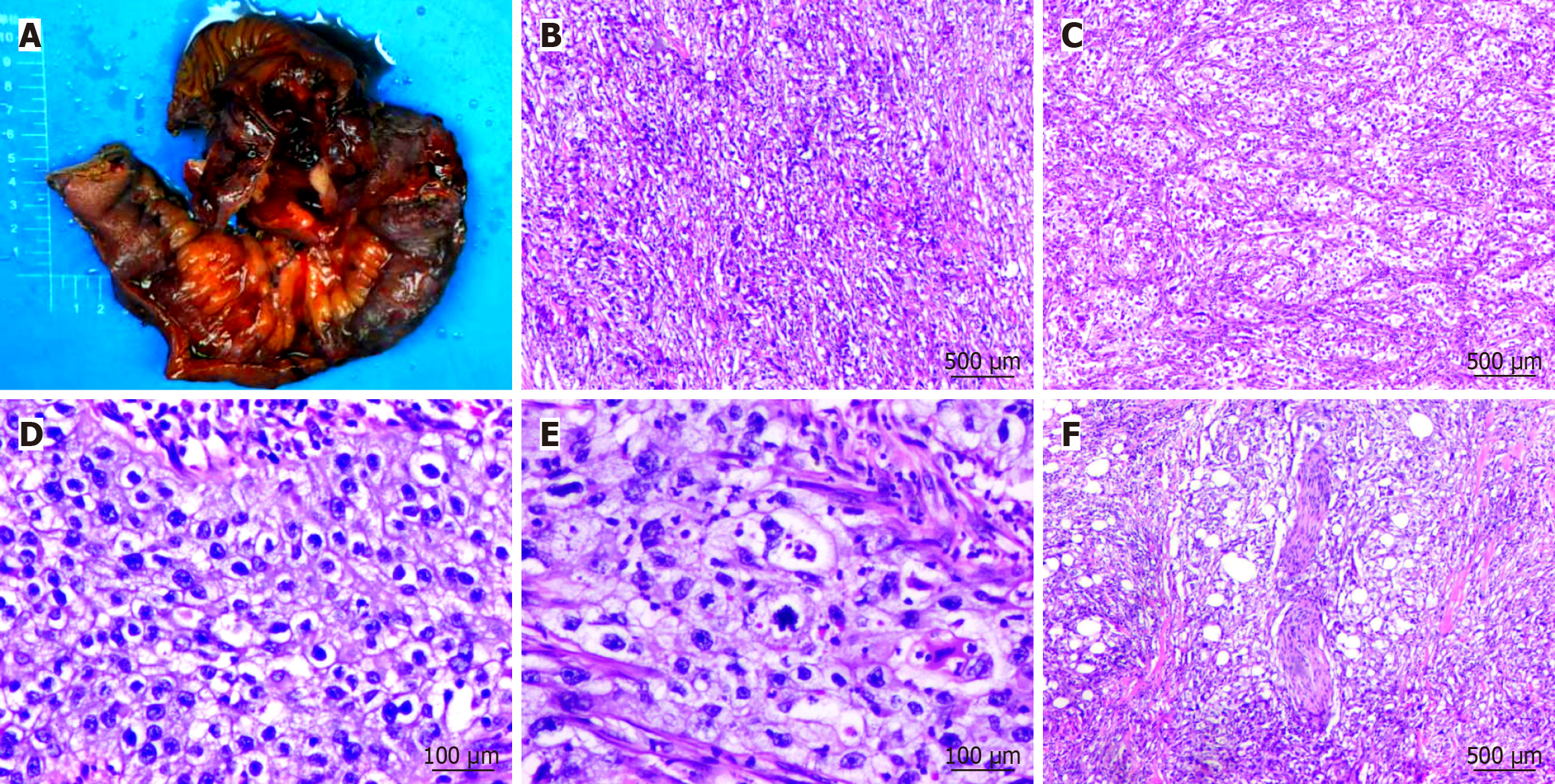
Figure 2 Macroscopic evaluation and hematoxylin-eosin staining of the small intestinal mass.
A: Gross specimen of primary ileal squamous cell carcinoma; B: Tumor cells arranged in solid sheet-like patterns (40 ×); C: Tumor cells forming nested clusters (40 ×); D: Tumor cells with abundant clear cytoplasm (200 ×); E: Pathological mitotic figures identified (200 ×); F: Evidence of perineural invasion by tumor cells (40 ×). The primary tumor mass observed in the resected intestinal specimen.
- Citation: Li QQ, Wei J, Fang LY, Zhou JL, Zhao HF. Primary ileal squamous cell carcinoma: A case report and review of literature. World J Clin Cases 2025; 13(36): 111835
- URL: https://www.wjgnet.com/2307-8960/full/v13/i36/111835.htm
- DOI: https://dx.doi.org/10.12998/wjcc.v13.i36.111835