
















©The Author(s) 2025.
World J Clin Cases. Dec 26, 2025; 13(36): 111835
Published online Dec 26, 2025. doi: 10.12998/wjcc.v13.i36.111835
Published online Dec 26, 2025. doi: 10.12998/wjcc.v13.i36.111835
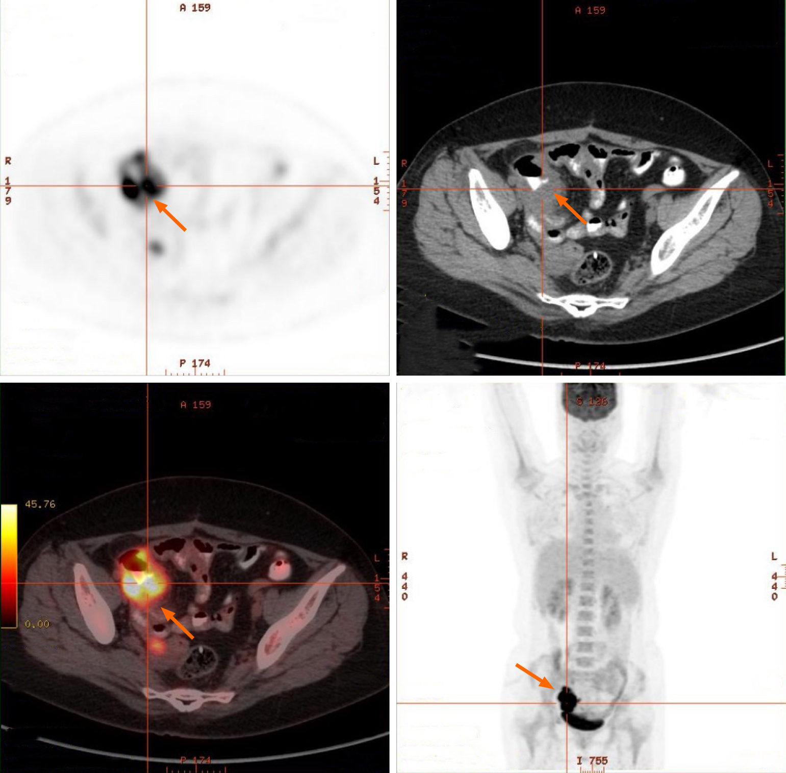
Figure 1 Small intestine positron emission tomography/computed tomography images.
Positron emission tomography/computed tomography revealed localized thickening of the small intestinal wall in the right pelvic region with increased metabolic activity, suggestive of implantation metastasis. The arrows point to the focal area of intestinal wall thickening with high metabolic activity.
- Citation: Li QQ, Wei J, Fang LY, Zhou JL, Zhao HF. Primary ileal squamous cell carcinoma: A case report and review of literature. World J Clin Cases 2025; 13(36): 111835
- URL: https://www.wjgnet.com/2307-8960/full/v13/i36/111835.htm
- DOI: https://dx.doi.org/10.12998/wjcc.v13.i36.111835