
















©The Author(s) 2025.
World J Clin Cases. Dec 16, 2025; 13(35): 115410
Published online Dec 16, 2025. doi: 10.12998/wjcc.v13.i35.115410
Published online Dec 16, 2025. doi: 10.12998/wjcc.v13.i35.115410
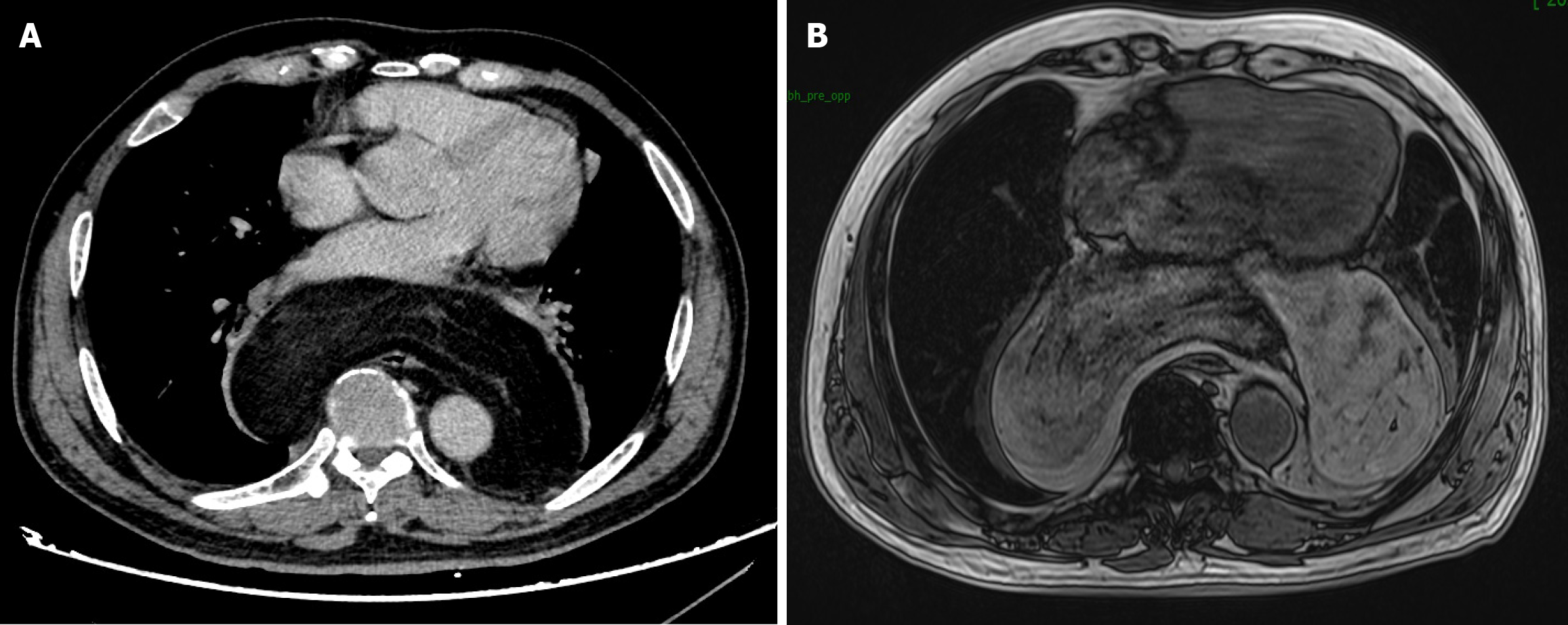
Figure 1 Enhanced computed tomography scan and chest enhanced magnetic resonance imaging.
A: Showing a giant posterior mediastinal tumor with a fatty component; B: Showing the tumor extensively involving the esophagus. There was still a small gap between the tumor and the esophagus.
- Citation: Ding YZ, Tang DQ, Zhao XJ. Esophageal fistula after resection of giant mediastinal liposarcoma: A case report. World J Clin Cases 2025; 13(35): 115410
- URL: https://www.wjgnet.com/2307-8960/full/v13/i35/115410.htm
- DOI: https://dx.doi.org/10.12998/wjcc.v13.i35.115410