
















©The Author(s) 2025.
World J Clin Cases. Dec 16, 2025; 13(35): 114672
Published online Dec 16, 2025. doi: 10.12998/wjcc.v13.i35.114672
Published online Dec 16, 2025. doi: 10.12998/wjcc.v13.i35.114672
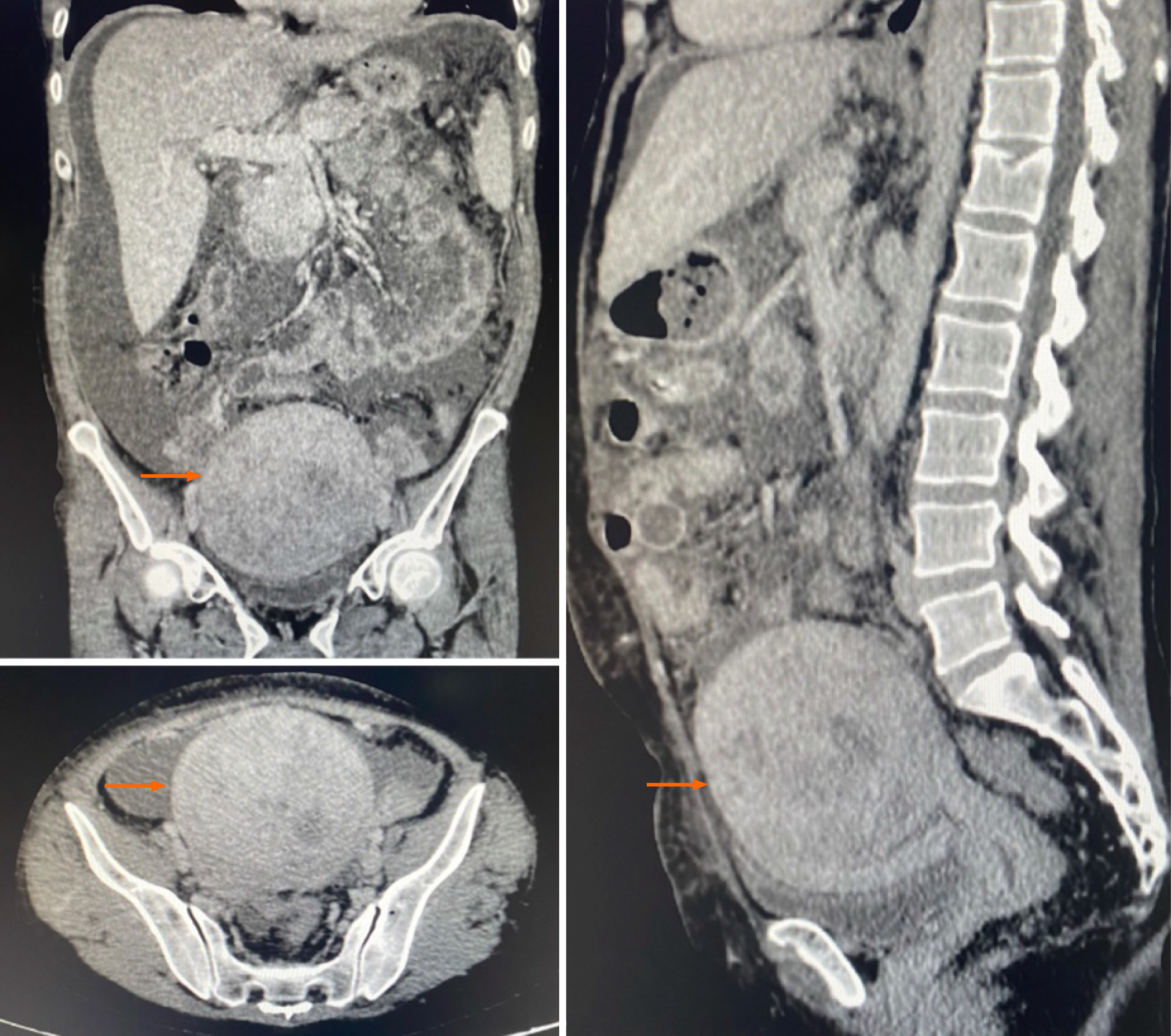
Figure 1 Computed tomography of the abdomen and pelvis showing a well-defined, heterogeneous nodular lesion located in the posterior wall/fundus of the uterus, measuring 10.
4 cm × 10.1 cm × 9.4 cm, associated with moderate ascites (orange arrow).
- Citation: Soares AABDS, Grillo TG, Almeida LC, Cavalleiro GST, Lopes MA, Craveiro MMS, Herrerias GSP, Baima JP, Saad-Hossne R, Sassaki LY. Endometrial stromal sarcoma in a patient with ulcerative colitis receiving immunosuppressive therapy: A case report and review of literature. World J Clin Cases 2025; 13(35): 114672
- URL: https://www.wjgnet.com/2307-8960/full/v13/i35/114672.htm
- DOI: https://dx.doi.org/10.12998/wjcc.v13.i35.114672