
















©The Author(s) 2025.
World J Clin Cases. Dec 6, 2025; 13(34): 114352
Published online Dec 6, 2025. doi: 10.12998/wjcc.v13.i34.114352
Published online Dec 6, 2025. doi: 10.12998/wjcc.v13.i34.114352
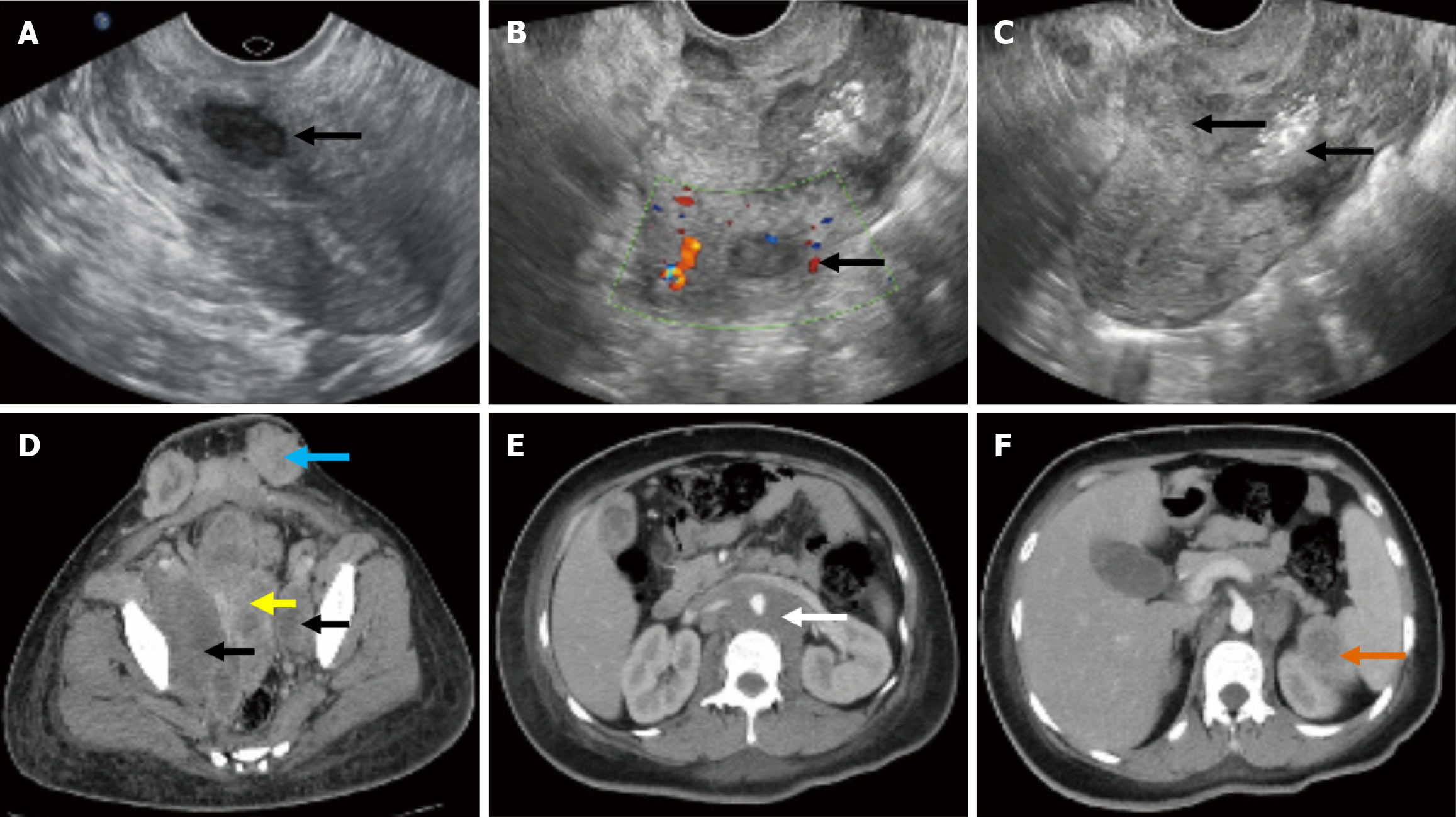
Figure 1 Transvaginal ultrasound images and enhanced computed tomography of the tumor and metastases.
A and B: The intramural uterine myoma located in the inferior-posterior wall of the uterus (A) 11 months before this pregnancy and (B) 13 days after chemotherapy (black arrow: The intramural uterine myoma); C: An uneven and weak-echo mass in the inferior uterine segment and cervical canal that invaded the entire layer of the muscle wall (black arrow: Tumor infiltration in the cervix ); D-F: Enhanced computed tomography of the tumor and metastases; yellow arrow: Tumor infiltration in the cervix and uterine cavity; black arrow: Tumor infiltration in the bilateral obturator lymph nodes; blue arrow: Tumor metastasis to subcutaneous tissue of the scar area of the abdominal wall; white arrow: Tumor metastasis to para-aortic lymph nodes; orange arrow: Tumor metastasis to spleen.
- Citation: Liu XJ, Wang P, Yang KX, Wang QL. Misdiagnosis and fatal outcome of advanced cervical adenosquamous carcinoma in pregnancy: A case report. World J Clin Cases 2025; 13(34): 114352
- URL: https://www.wjgnet.com/2307-8960/full/v13/i34/114352.htm
- DOI: https://dx.doi.org/10.12998/wjcc.v13.i34.114352