
















©The Author(s) 2025.
World J Clin Cases. Dec 6, 2025; 13(34): 110925
Published online Dec 6, 2025. doi: 10.12998/wjcc.v13.i34.110925
Published online Dec 6, 2025. doi: 10.12998/wjcc.v13.i34.110925
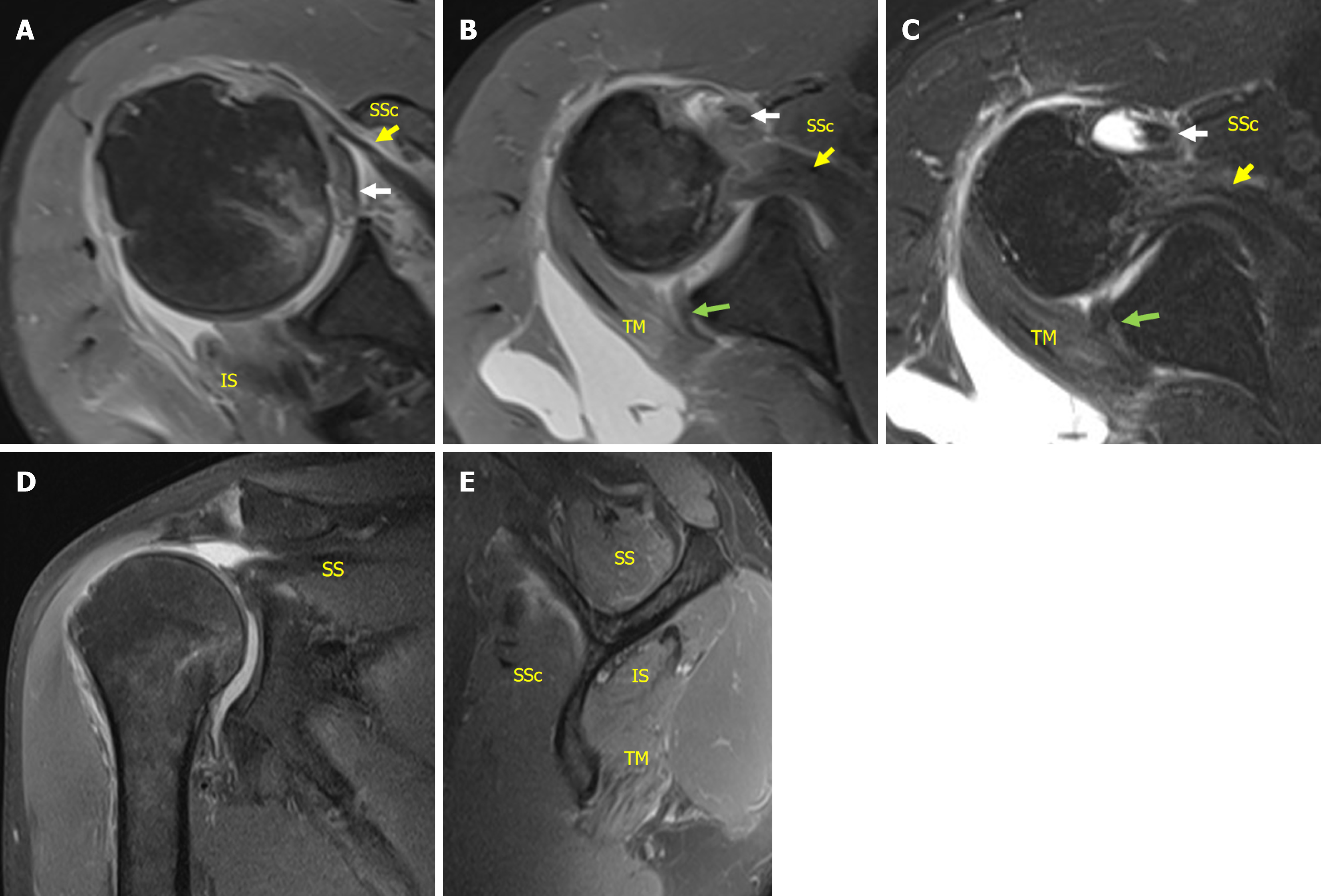
Figure 3 Magnetic resonance imaging performed 2 weeks after the injury.
A-C: Images revealed complete tear of infraspinatus tendon and an upper two-thirds complete tear of the subscapularis tendon (LaFosse stage 3) as well as medial dislocation and rupture of the long head of the biceps tendon. The posterior-inferior labrum was torn but remained attached to the scapular periosteum and partially to the glenoid. Glenoid bone loss was minimal; D: Complete tear of supraspinatus tendon with retraction to the glenoid (Patte stage 3). The rotator cuff tear was classified as the most severe topographic type (involved segment 6 in the sagittal plane); E: Fatty infiltration was mild (Goutallier grade, supraspinatus/infraspinatus/subscapularis/teres minor: 2/1/1/0). White arrow: Long head of the biceps tendon; Yellow arrow: Subscapularis; Green arrow: Posterior-inferior labrum; SSc: Subscapularis; SS: Supraspinatus; IS: Infraspinatus; TM: Teres minor.
- Citation: Liu MY, Lin CH, Chen SH, Ding YS, Chiang CH. Acute massive rotator cuff tear and biceps tendon dislocation following posterior shoulder dislocation: A case report. World J Clin Cases 2025; 13(34): 110925
- URL: https://www.wjgnet.com/2307-8960/full/v13/i34/110925.htm
- DOI: https://dx.doi.org/10.12998/wjcc.v13.i34.110925