
















©The Author(s) 2025.
World J Clin Cases. Nov 26, 2025; 13(33): 113312
Published online Nov 26, 2025. doi: 10.12998/wjcc.v13.i33.113312
Published online Nov 26, 2025. doi: 10.12998/wjcc.v13.i33.113312
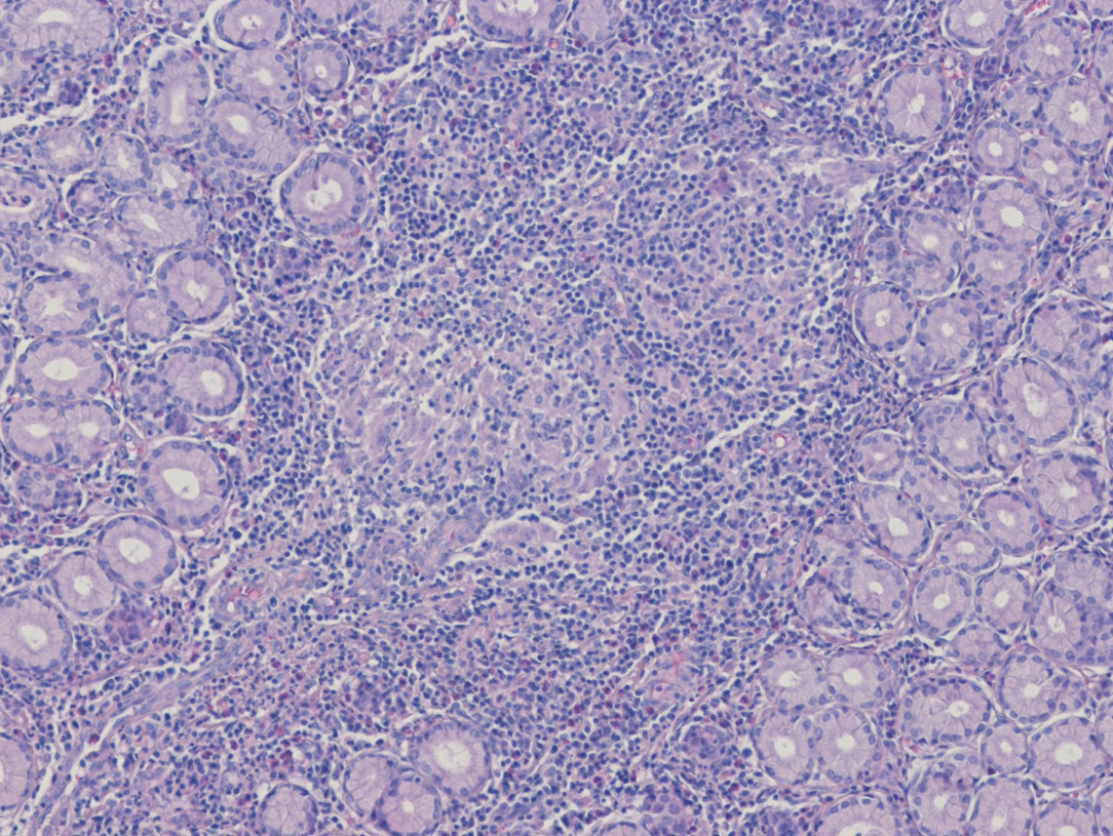
Figure 2 Histologic findings of a gastric subepithelial tumor-like lesion.
Histopathological image showing a well-formed non-caseating granuloma composed of epithelioid histiocytes and surrounding lymphocytes, with marked infiltration of chronic inflammatory cells including plasma cells, findings consistent with upper gastrointestinal involvement in Crohn’s disease. Adjacent normal gastric glands are also observed (hematoxylin and eosin stain, × 100).
- Citation: Park YE. Gastric Crohn’s disease presenting as a subepithelial tumor: A case report. World J Clin Cases 2025; 13(33): 113312
- URL: https://www.wjgnet.com/2307-8960/full/v13/i33/113312.htm
- DOI: https://dx.doi.org/10.12998/wjcc.v13.i33.113312