
















©The Author(s) 2025.
World J Clin Cases. Nov 26, 2025; 13(33): 113312
Published online Nov 26, 2025. doi: 10.12998/wjcc.v13.i33.113312
Published online Nov 26, 2025. doi: 10.12998/wjcc.v13.i33.113312
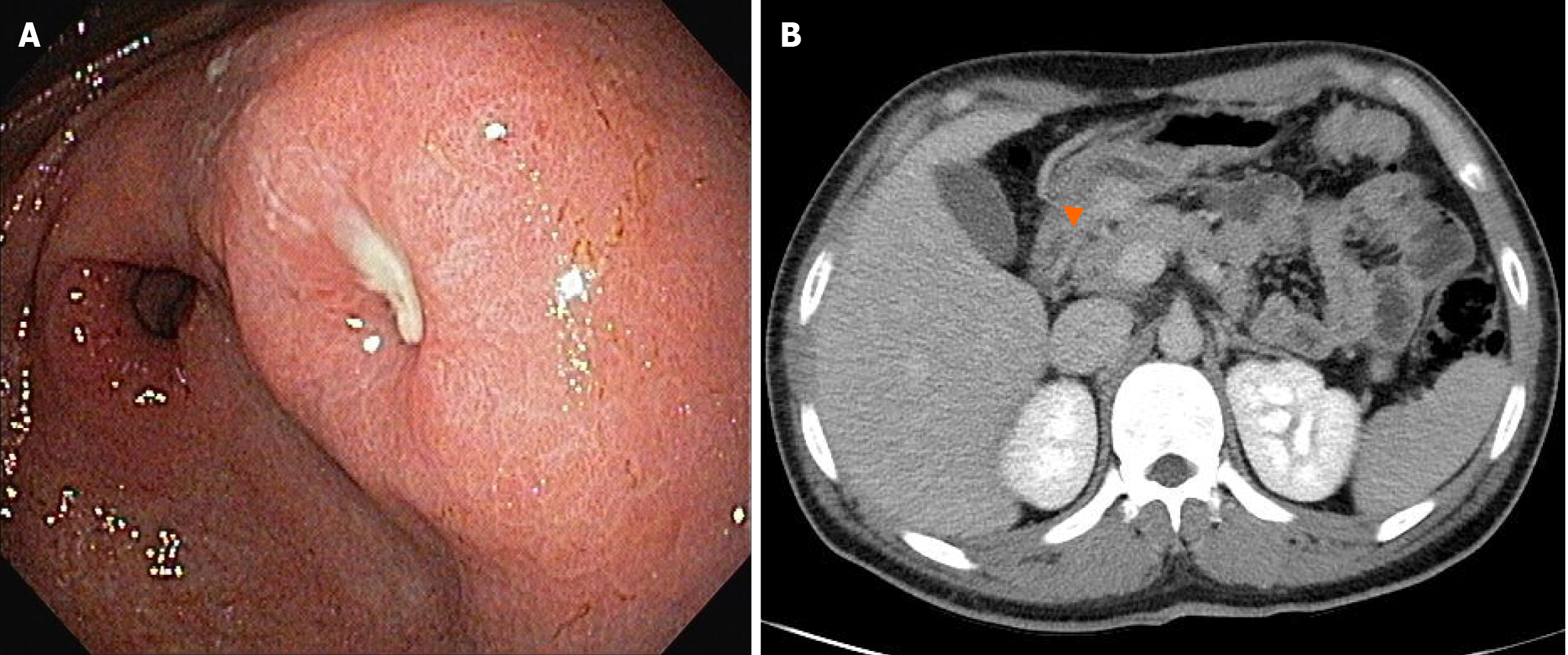
Figure 1 Endoscopy and abdominal computed tomography findings.
A: Endoscopic image showing a protruding lesion with central ulceration on the posterior wall of the distal gastric antrum; B: Axial contrast-enhanced computed tomography image showing a well-defined, hypervascular subepithelial lesion (orange arrowhead) along the posterior wall of the distal gastric antrum.
- Citation: Park YE. Gastric Crohn’s disease presenting as a subepithelial tumor: A case report. World J Clin Cases 2025; 13(33): 113312
- URL: https://www.wjgnet.com/2307-8960/full/v13/i33/113312.htm
- DOI: https://dx.doi.org/10.12998/wjcc.v13.i33.113312