
















©The Author(s) 2025.
World J Clin Cases. Nov 26, 2025; 13(33): 113216
Published online Nov 26, 2025. doi: 10.12998/wjcc.v13.i33.113216
Published online Nov 26, 2025. doi: 10.12998/wjcc.v13.i33.113216
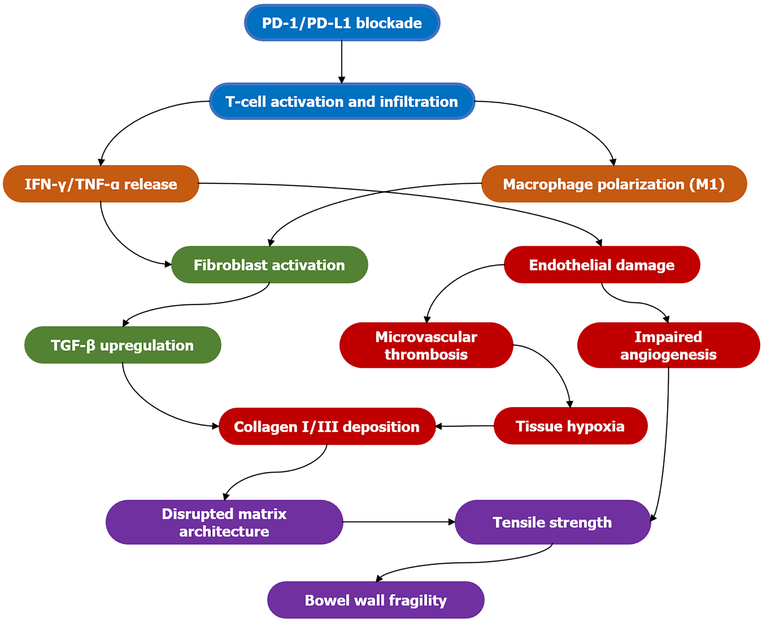
Figure 1 Pathogenesis of immune-mediated bowel wall remodeling.
Stage 1 (blue): Immune checkpoint inhibition is lifted, activating and infiltrating T cells; stage 2 (orange): Activated T cells and M1-type macrophages release large amounts of interferon-gamma and tumor necrosis factor alpha; stage 3 (green): Fibroblasts are activated and upregulate transforming growth factor beta, initiating the fibrosis program; stage 4 (red): Transforming growth factor beta promotes the deposition of type I/III collagen; simultaneously, inflammatory mediators cause vascular endothelial damage, microthrombosis formation, tissue hypoxia, and impaired angiogenesis; stage 5 (purple): Disordered collagen deposition disrupts the matrix structure, leading to reduced tissue tension and ultimately increased intestinal wall fragility. PD: Programmed death; IFN-γ: Interferon-gamma; TNF-α: Tumor necrosis factor alpha; TGF-β: Transforming growth factor beta.
- Citation: Wang RG. When new therapies arrive, we should think more: The unseen challenges of immunotherapy-induced remodeling. World J Clin Cases 2025; 13(33): 113216
- URL: https://www.wjgnet.com/2307-8960/full/v13/i33/113216.htm
- DOI: https://dx.doi.org/10.12998/wjcc.v13.i33.113216