
















©The Author(s) 2025.
World J Clin Cases. Nov 16, 2025; 13(32): 111879
Published online Nov 16, 2025. doi: 10.12998/wjcc.v13.i32.111879
Published online Nov 16, 2025. doi: 10.12998/wjcc.v13.i32.111879
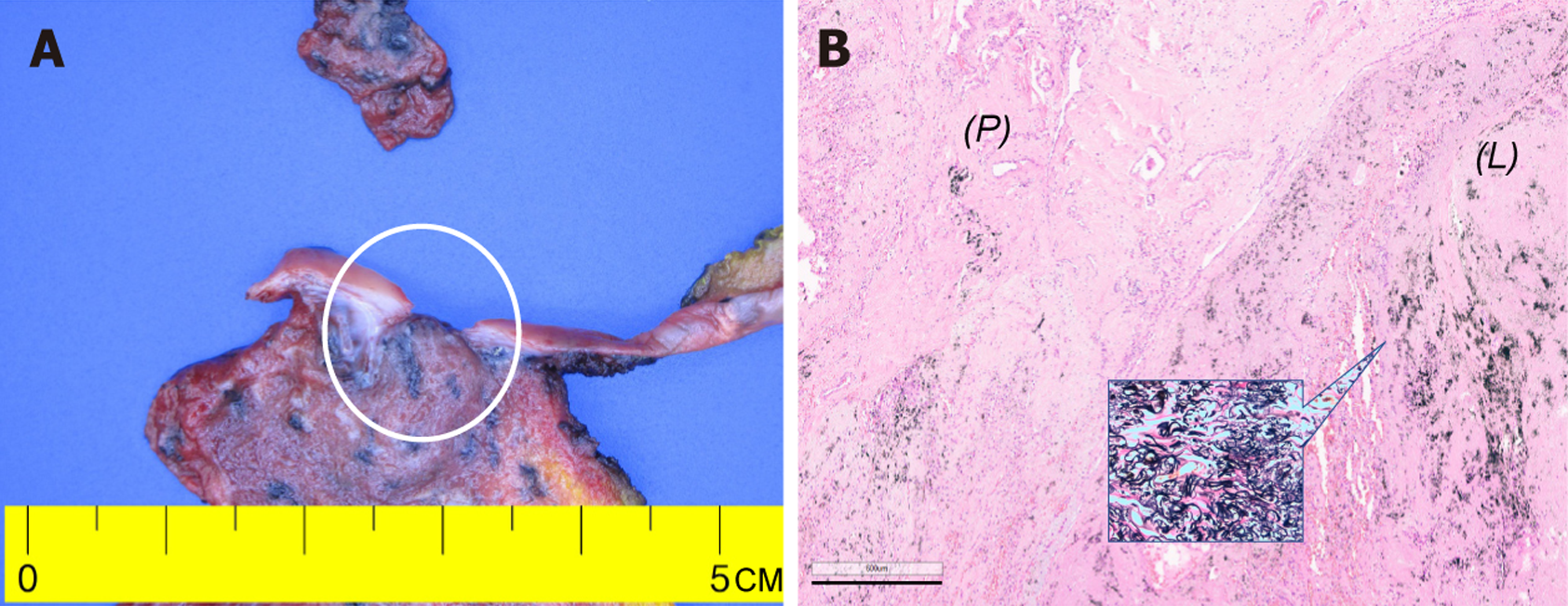
Figure 2 Gross and histopathology of the resected left upper lobe consistent with pleuroparenchymal fibroelastosis.
A: Gross specimen with diffuse visceral pleural thickening and a firm gray to black subpleural lesion (white circle) corresponding to the radiological abnormality, scale: Centimeter ruler provided; B: Hematoxylin and eosin staining (× 40 magnification) showed dense fibroelastic thickening of the visceral pleura extending into the adjacent subpleural lung parenchyma with minimal inflammation and no malignant features, inset: Elastic Van Gieson staining (× 100 magnification) highlighted abundant elastic fiber deposition within the pleura and subpleural alveolar septa, consistent with pleuroparenchymal fibroelastosis (scale bar: 600 μm). L: Lung parenchyma; P: Pleura.
- Citation: Jung HS, Kim HJ, Kim KW. Early pleuroparenchymal fibroelastosis mimicking lung malignancy: A case report. World J Clin Cases 2025; 13(32): 111879
- URL: https://www.wjgnet.com/2307-8960/full/v13/i32/111879.htm
- DOI: https://dx.doi.org/10.12998/wjcc.v13.i32.111879