
















©The Author(s) 2025.
World J Clin Cases. Nov 16, 2025; 13(32): 111879
Published online Nov 16, 2025. doi: 10.12998/wjcc.v13.i32.111879
Published online Nov 16, 2025. doi: 10.12998/wjcc.v13.i32.111879
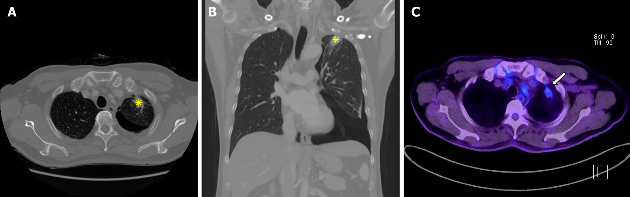
Figure 1 Imaging results.
A and B: Chest computed tomography demonstrated left upper lobe subpleural consolidation with pleural thickening and associated pneumothorax. Axial chest computed tomography showed subpleural consolidation (asterisks) and pleural thickening in the left upper lobe (A). Coronal chest computed tomography confirmed subpleural consolidation (asterisks) with adjacent pneumothorax (B); C: 18F-fluorodeoxyglucose positron emission tomography/computed tomography. Moderate fluorodeoxyglucose uptake (maximum standardized uptake value = 3.2) was observed in the left upper lobe (arrows), suggesting malignancy.
- Citation: Jung HS, Kim HJ, Kim KW. Early pleuroparenchymal fibroelastosis mimicking lung malignancy: A case report. World J Clin Cases 2025; 13(32): 111879
- URL: https://www.wjgnet.com/2307-8960/full/v13/i32/111879.htm
- DOI: https://dx.doi.org/10.12998/wjcc.v13.i32.111879