
















©The Author(s) 2025.
World J Clin Cases. Nov 16, 2025; 13(32): 111796
Published online Nov 16, 2025. doi: 10.12998/wjcc.v13.i32.111796
Published online Nov 16, 2025. doi: 10.12998/wjcc.v13.i32.111796
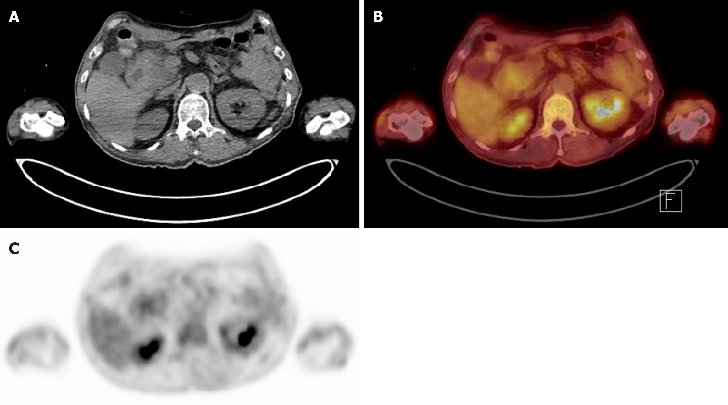
Figure 4 Positron emission tomography-computed tomography images.
A: Axial computed tomography torso image. Mass at periampullary area; B: Fusion positron emission tomography-computed tomography image demonstrating a mild hypermetabolic mass-like lesion in the second portion of the duodenum near the ampulla of Vater of unknown clinical significance; C: Axial positron emission tomography image showing the corresponding focal uptake in the same region of (B).
- Citation: Kim N, Lee H, Park D. Challenging diagnosis of groove pancreatitis with severe gastric outlet obstruction: A case report. World J Clin Cases 2025; 13(32): 111796
- URL: https://www.wjgnet.com/2307-8960/full/v13/i32/111796.htm
- DOI: https://dx.doi.org/10.12998/wjcc.v13.i32.111796