
















©The Author(s) 2025.
World J Clin Cases. Nov 16, 2025; 13(32): 111796
Published online Nov 16, 2025. doi: 10.12998/wjcc.v13.i32.111796
Published online Nov 16, 2025. doi: 10.12998/wjcc.v13.i32.111796
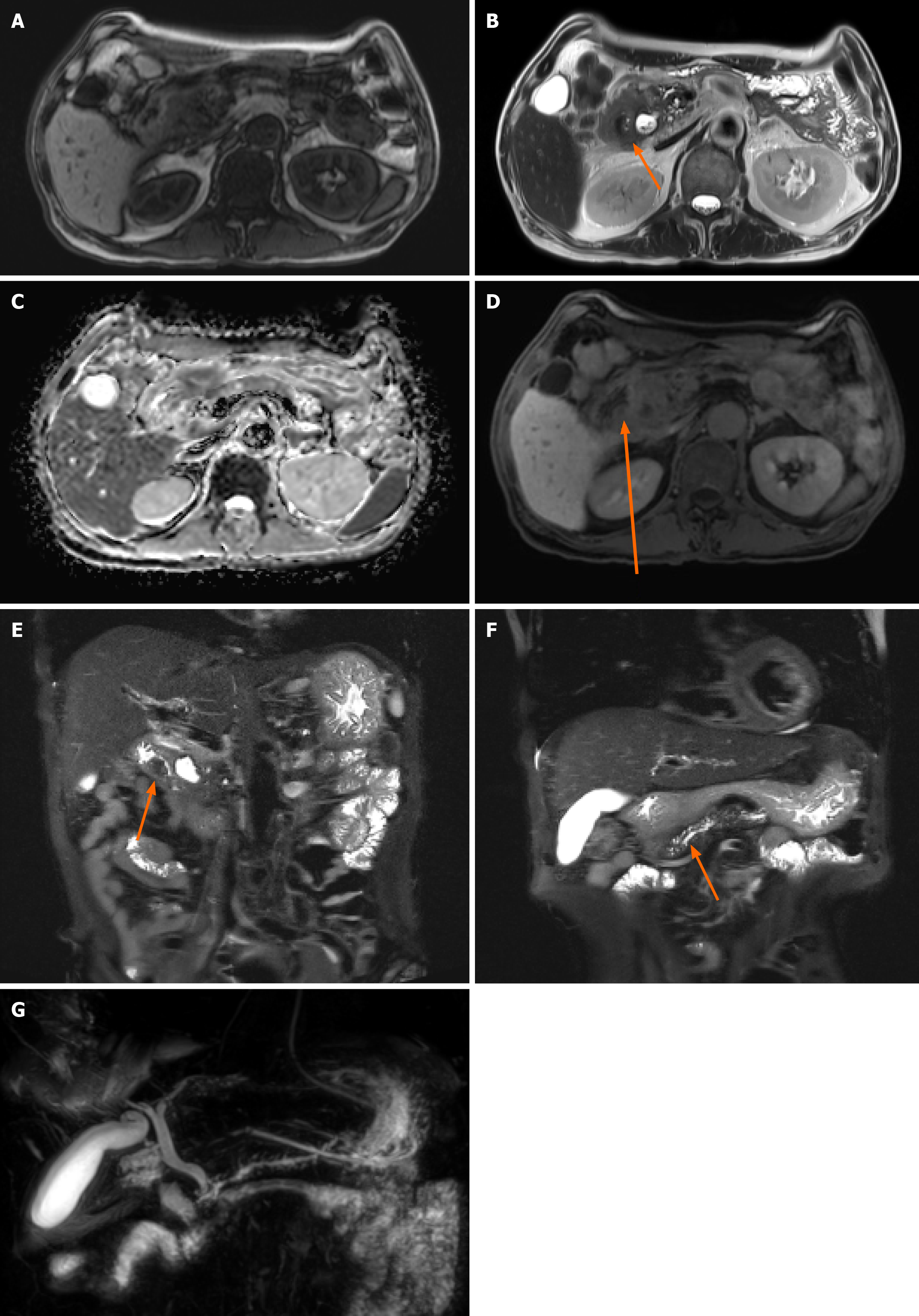
Figure 2 Pancreatic magnetic resonance imaging findings.
A: T1 weighted image; B: T2 weighted axial image; C: Diffusion weighted image; D: Vibe image; E and F: T2 weight coronal image; G: Magnetic resonance cholangiopancreatography. Magnetic resonance imaging showing a lesion in the ampulla of Vater with no clear T1 weighted and diffusion restriction image (A and C) but prominent enhancement, which is suspicious for ampulla of Vater cancer (B, D and E); magnetic resonance imaging showing main pancreatic duct upstream dilatation (F and G).
- Citation: Kim N, Lee H, Park D. Challenging diagnosis of groove pancreatitis with severe gastric outlet obstruction: A case report. World J Clin Cases 2025; 13(32): 111796
- URL: https://www.wjgnet.com/2307-8960/full/v13/i32/111796.htm
- DOI: https://dx.doi.org/10.12998/wjcc.v13.i32.111796