
















©The Author(s) 2025.
World J Clin Cases. Nov 16, 2025; 13(32): 111525
Published online Nov 16, 2025. doi: 10.12998/wjcc.v13.i32.111525
Published online Nov 16, 2025. doi: 10.12998/wjcc.v13.i32.111525
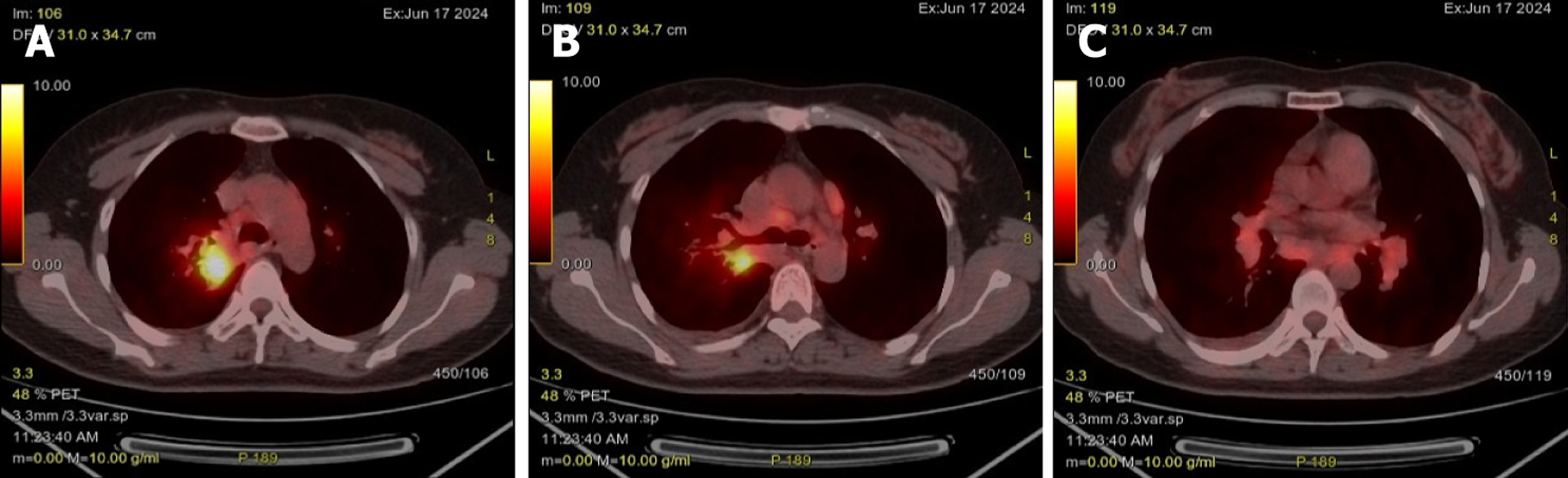
Figure 3 Reveals a hypermetabolic consolidation in the right upper lung lobe with multiple fluorodeoxyglucose-avid enlarged mediastinal and hilar lymph nodes, initially suggestive of primary lung malignancy with lymphangitic carcinomatosis and nodal metastases.
A and B: Hypermetabolic consolidation in the right lung; C: Multiple enlarged lymph nodes in the mediastinum and hilar regions.
- Citation: Yu WX, Zhan FF, Hong PY, Huang MH, Chen YY, Lin YL, Zhang XB. Atypical sarcoidosis diagnosed using endobronchial ultrasound-guided mediastinal lymph node biopsy with fine biopsy forceps: Two case reports. World J Clin Cases 2025; 13(32): 111525
- URL: https://www.wjgnet.com/2307-8960/full/v13/i32/111525.htm
- DOI: https://dx.doi.org/10.12998/wjcc.v13.i32.111525