
















©The Author(s) 2025.
World J Clin Cases. Nov 16, 2025; 13(32): 110897
Published online Nov 16, 2025. doi: 10.12998/wjcc.v13.i32.110897
Published online Nov 16, 2025. doi: 10.12998/wjcc.v13.i32.110897
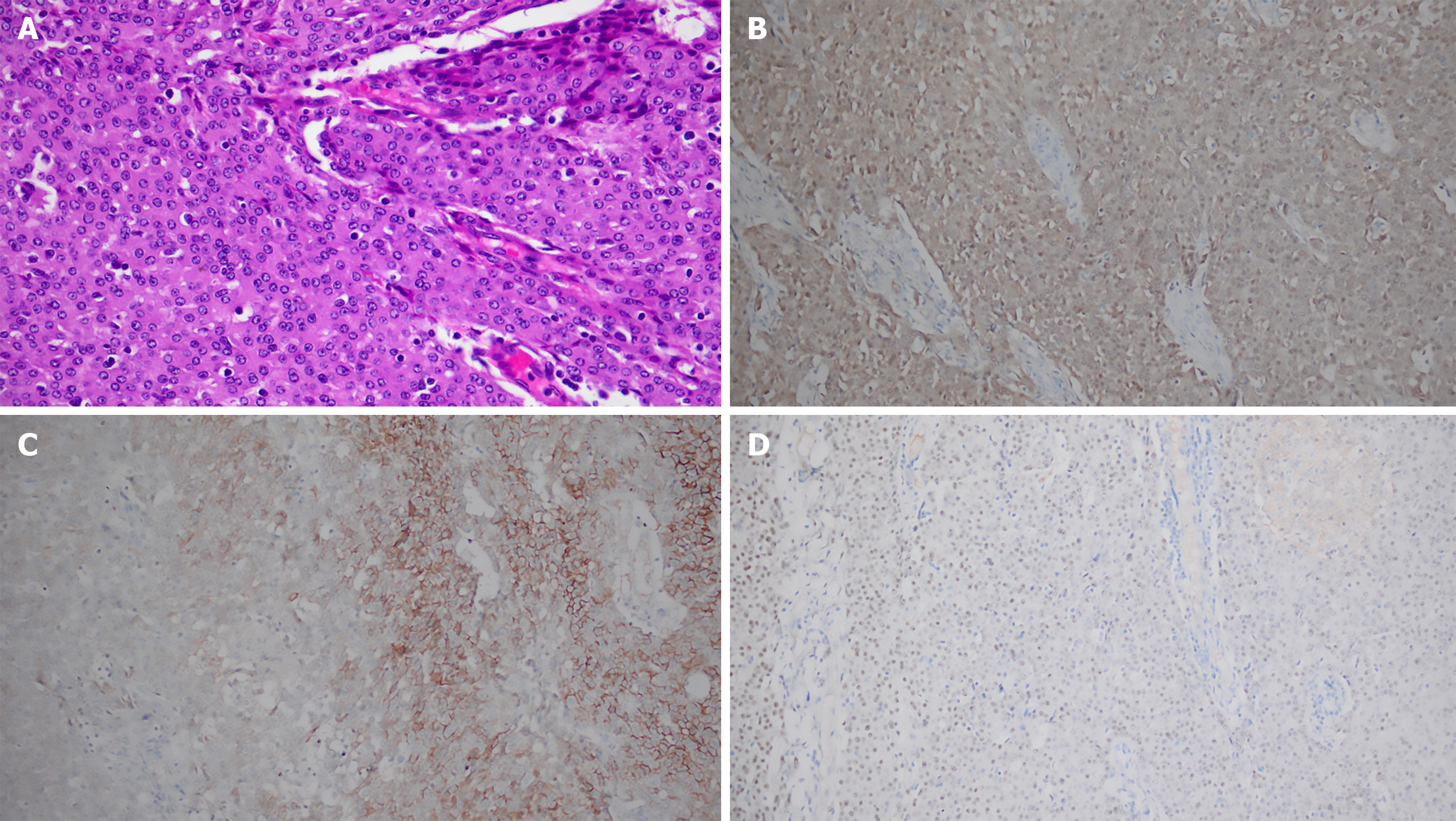
Figure 4 Histopathological analysis of the resected specimen.
A: Hematoxylin and eosin (HE) staining (× 400): Epithelioid tumor cells arranged in sheets, exhibiting large cell volume, eosinophilic cytoplasm, and marked nuclear atypia; B-D: Immunohistochemistry (IHC) (× 200); B: Calretinin: Strong and diffuse cytoplasmic/nuclear positivity in tumor cells; C: D2-40: Partial membranous positivity in tumor cells; D: WT-1: Focal nuclear positivity in tumor cells. The combined morphological and IHC profile (Calretinin+/CK5/6+/WT-1 focal+/TTF-1–) supports the diagnosis of epithelioid malignant pleural mesothelioma.
- Citation: Aisikaer A, Sun MM, Shen J. Positron emission tomography/computed tomography in risk-factor-negative young female with malignant pleural mesothelioma: A case report and review of literature. World J Clin Cases 2025; 13(32): 110897
- URL: https://www.wjgnet.com/2307-8960/full/v13/i32/110897.htm
- DOI: https://dx.doi.org/10.12998/wjcc.v13.i32.110897