
















©The Author(s) 2025.
World J Clin Cases. Nov 16, 2025; 13(32): 110813
Published online Nov 16, 2025. doi: 10.12998/wjcc.v13.i32.110813
Published online Nov 16, 2025. doi: 10.12998/wjcc.v13.i32.110813
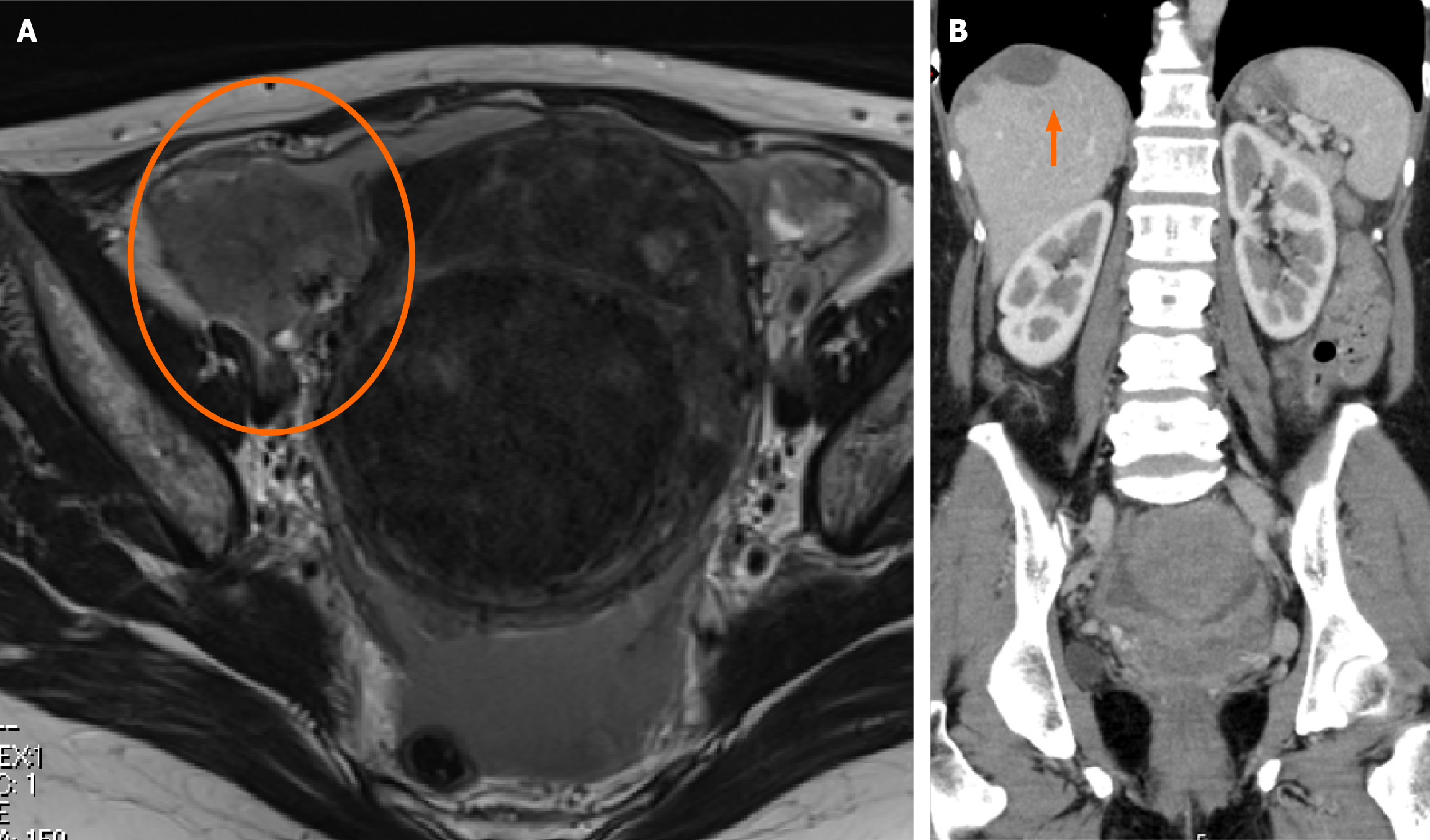
Figure 3 Image findings.
A: Plain magnetic resonance imaging T2-weighted image: 7-cm mass is noted in the right adnexa; B: Contrast-enhanced computed tomography, with findings suggestive of diffuse peritoneal dissemination from the pelvis to the diaphragm. The arrow indicates the subdiaphragmatic disseminated lesion.
- Citation: Sagawa S, Ito F, Nakatani M, Kurose S, Niiro E, Taniguchi M, Toyoda S, Sado T, Morita K. Mesonephric-like adenocarcinoma presenting with malignant peritonitis and suspected to originate from the fallopian tube: A case report. World J Clin Cases 2025; 13(32): 110813
- URL: https://www.wjgnet.com/2307-8960/full/v13/i32/110813.htm
- DOI: https://dx.doi.org/10.12998/wjcc.v13.i32.110813