
















©The Author(s) 2025.
World J Clin Cases. Nov 6, 2025; 13(31): 112023
Published online Nov 6, 2025. doi: 10.12998/wjcc.v13.i31.112023
Published online Nov 6, 2025. doi: 10.12998/wjcc.v13.i31.112023
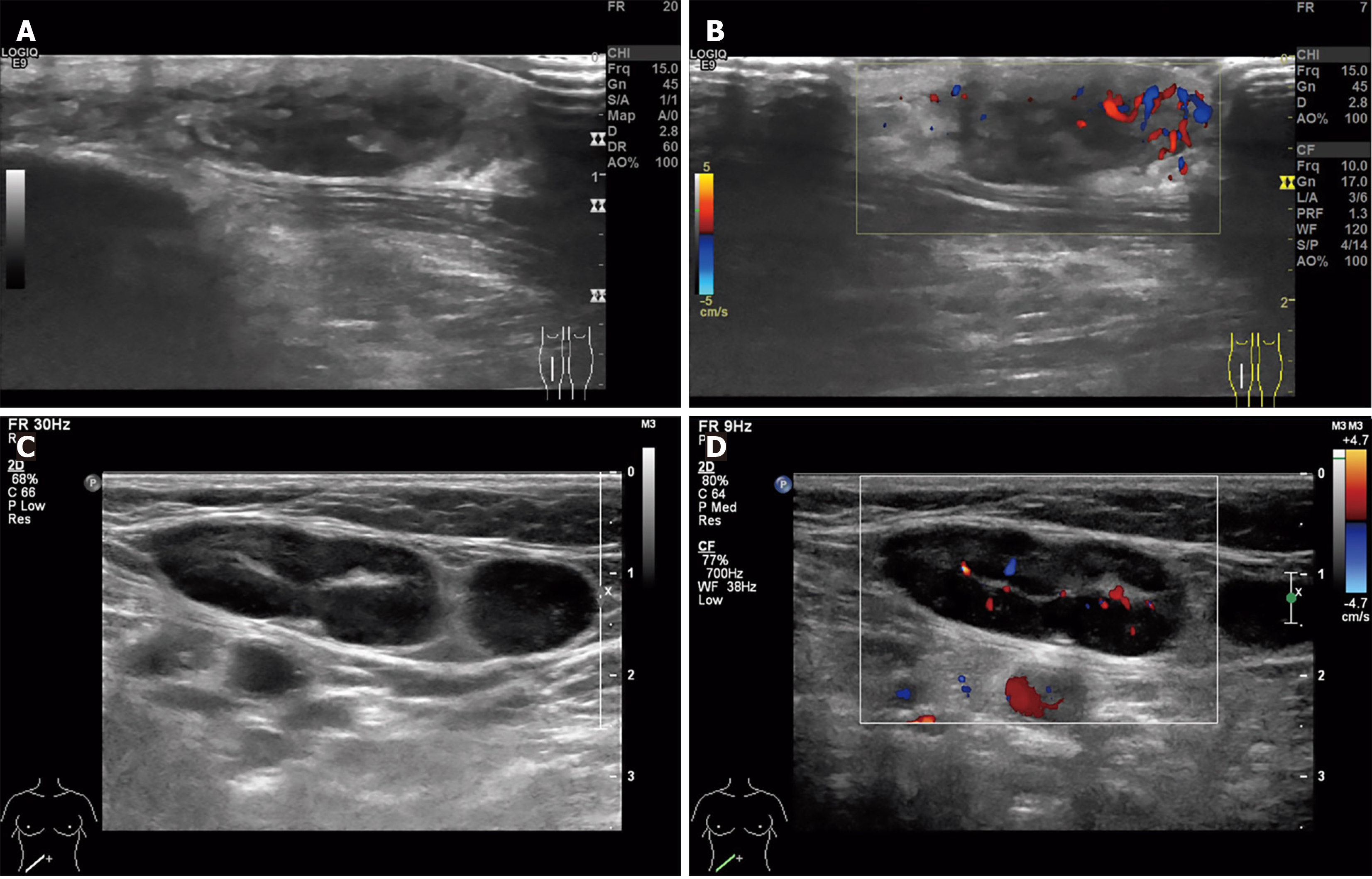
Figure 2 Ultrasound imaging of the right lower leg mass and right inguinal lymph nodes.
A: A hypoechoic subcutaneous mass (25 mm × 9 mm × 25 mm) in the mid-right lower leg, exhibiting irregular margins and an ill-defined shape; B: The adjacent tissue demonstrates increased echogenicity, accompanied by prominent peripheral vascularity; C: Multiple enlarged lymph nodes are observed in the right inguinal region, with the largest measuring 30 mm × 13 mm and showing asymmetrical cortical thickening; D: Intranodal blood flow appears as punctate/Linear vascular signals.
- Citation: Zhou PY, Chen W, Wang L. Mantle cell lymphoma presenting primarily as cutaneous lesions: A case report. World J Clin Cases 2025; 13(31): 112023
- URL: https://www.wjgnet.com/2307-8960/full/v13/i31/112023.htm
- DOI: https://dx.doi.org/10.12998/wjcc.v13.i31.112023