
















©The Author(s) 2025.
World J Clin Cases. Nov 6, 2025; 13(31): 110624
Published online Nov 6, 2025. doi: 10.12998/wjcc.v13.i31.110624
Published online Nov 6, 2025. doi: 10.12998/wjcc.v13.i31.110624
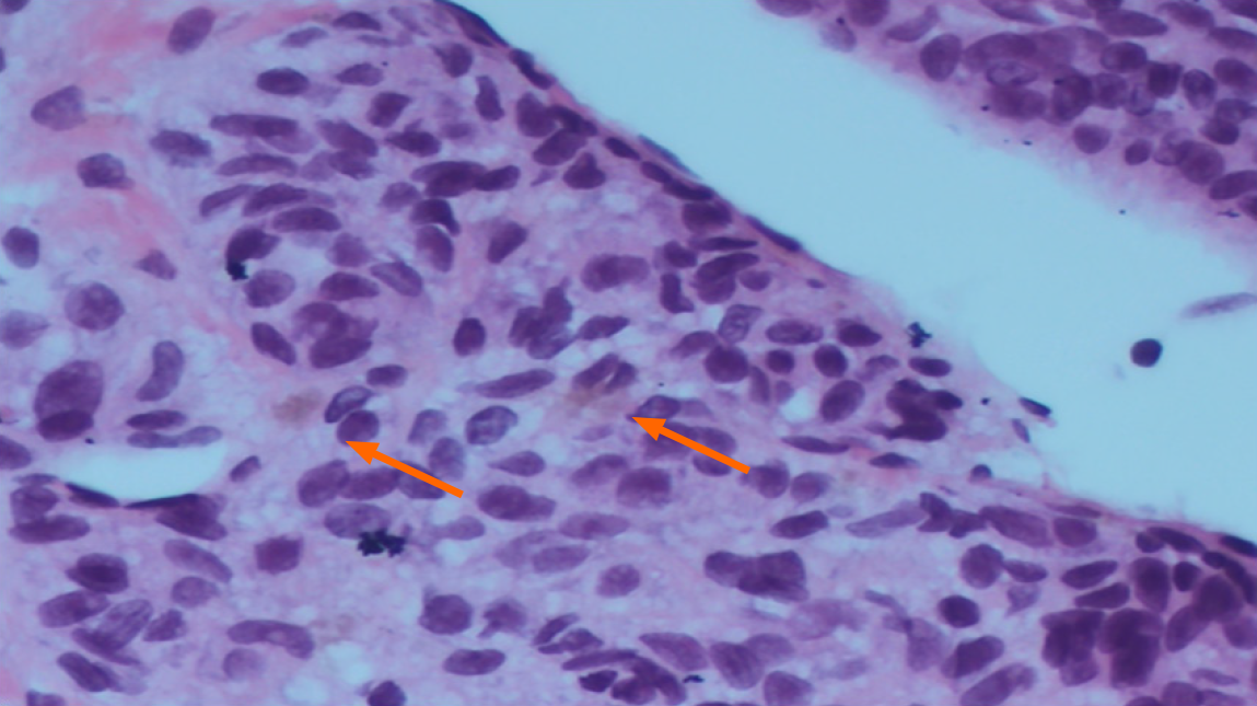
Figure 2 Microscopic view shows a sparse amount of normal liver parenchyma, with the remainder of the biopsy consisting of a necrotic tumor infiltrate composed of clusters and short strands of atypical, oval to spindle-shaped cells, with polymorphic nuclei and prominent nucleoli.
Arrows are pointing to the tumor cells.
- Citation: Domislovic V, Sesa V, Kosuta I, Bulimbasic S, Mrzljak A. Liver failure due to metastatic melanoma: A case report. World J Clin Cases 2025; 13(31): 110624
- URL: https://www.wjgnet.com/2307-8960/full/v13/i31/110624.htm
- DOI: https://dx.doi.org/10.12998/wjcc.v13.i31.110624