
















©The Author(s) 2025.
World J Clin Cases. Nov 6, 2025; 13(31): 109584
Published online Nov 6, 2025. doi: 10.12998/wjcc.v13.i31.109584
Published online Nov 6, 2025. doi: 10.12998/wjcc.v13.i31.109584
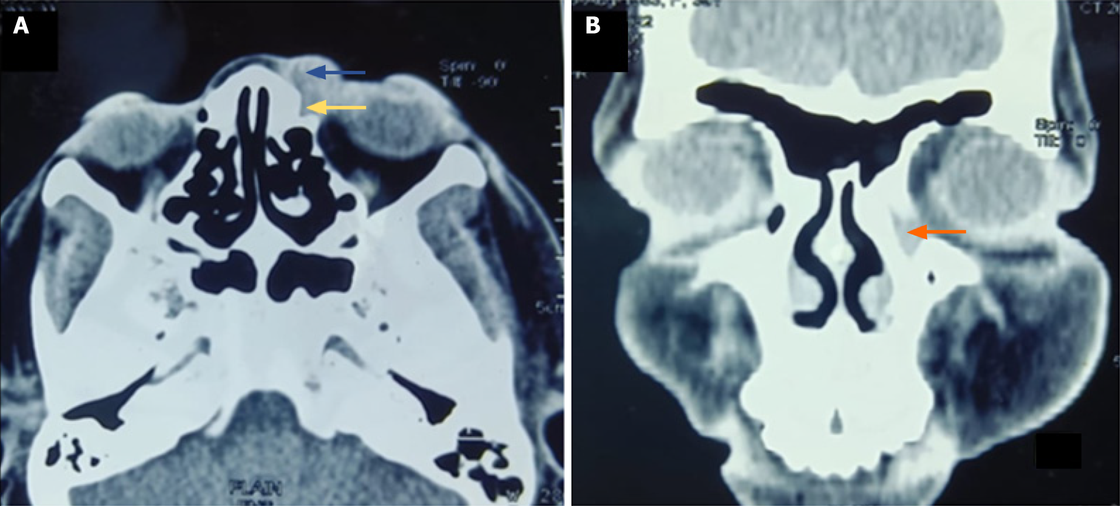
Figure 3 Computed tomography.
A and B: Isodense mass lesion in lacrimal sac area showing extension into preseptal area (marked with yellow arrow), left nasolacrimal duct (marked with orange arrow).
- Citation: Panda BB, Kar A, Koppalu Lingaraju T, Ayyanar P. Acquired cutaneous fistula in the periocular area: A case report. World J Clin Cases 2025; 13(31): 109584
- URL: https://www.wjgnet.com/2307-8960/full/v13/i31/109584.htm
- DOI: https://dx.doi.org/10.12998/wjcc.v13.i31.109584