
















©The Author(s) 2025.
World J Clin Cases. Oct 26, 2025; 13(30): 111020
Published online Oct 26, 2025. doi: 10.12998/wjcc.v13.i30.111020
Published online Oct 26, 2025. doi: 10.12998/wjcc.v13.i30.111020
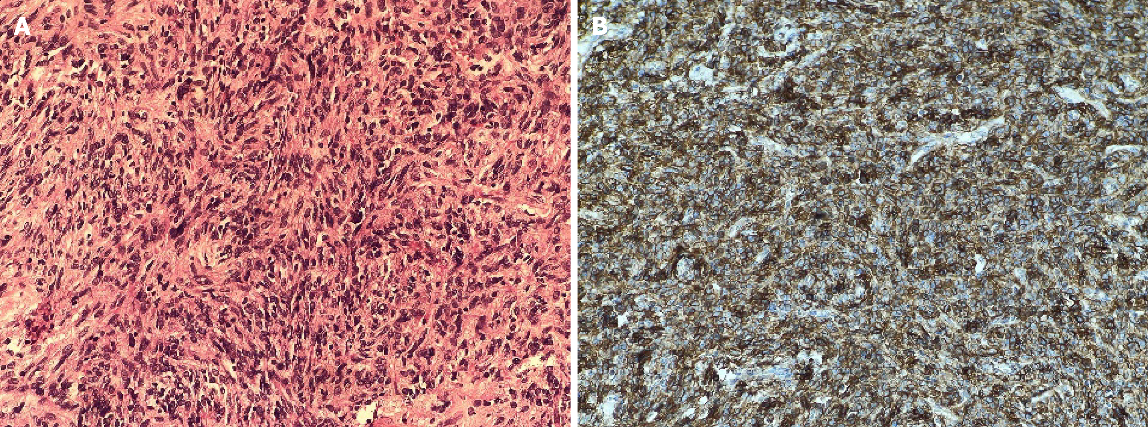
Figure 2 Histopathological characteristics of the excised specimen confirming the diagnosis of dermatofibrosarcoma protruberans.
A: Histopathology hematoxylin and eosin staining, 400 ×. The tumor is composed of spindle cells arranged in a storiform pattern. The cells showed elongated nuclei, abundant fibrillar cytoplasm, and mild nuclear atypia; B: Immunohistochemistry showed that the tumor cells have strong and diffuse positivity for cluster of differentiation 34.
- Citation: Panda BB, Gunasekar S, Agarwal U, Koppalu Lingaraju T, Adhya AK. Recurrent dermatofibrosarcoma protuberans involving the lacrimal sac: A case report. World J Clin Cases 2025; 13(30): 111020
- URL: https://www.wjgnet.com/2307-8960/full/v13/i30/111020.htm
- DOI: https://dx.doi.org/10.12998/wjcc.v13.i30.111020