
















©The Author(s) 2025.
World J Clin Cases. Oct 26, 2025; 13(30): 110475
Published online Oct 26, 2025. doi: 10.12998/wjcc.v13.i30.110475
Published online Oct 26, 2025. doi: 10.12998/wjcc.v13.i30.110475
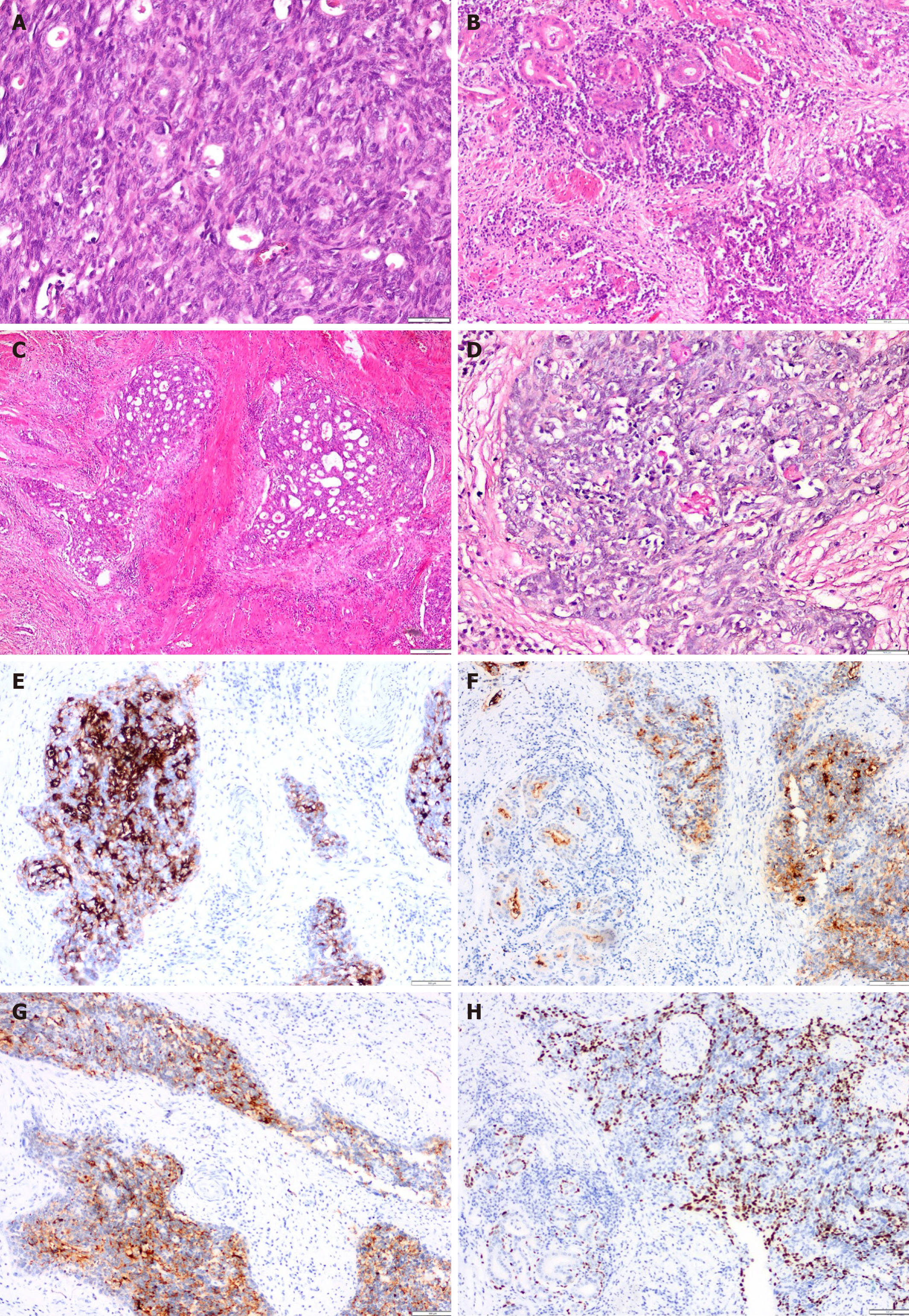
Figure 2 Microscopic appearance of the tumor.
A: The tumor was primarily composed of solid tumor islands. These areas comprised squamous epithelium, intermediate, and adenoid cells with larger cytoplasm and occasional epithelial characteristics; B: Areas displaying gland-like structures (notably in the upper left) were adjacent to solid regions (located in the lower right); C: In another area the glandular structures were interspersed among solid regions, suggesting a mucoepidermoid morphology; D: The mucicarmine staining technique revealed prominent pink staining of mucin (MUC) within the luminal spaces of the glandular structures; E: In these areas, the epithelial cells forming gland-like structures demonstrated MUC1 positivity, contrasting with the surrounding squamous epithelial cells that exhibited no staining; F: Carcinoembryonic antigen positivity was noted in the adenoid areas in both the lumen and the cytoplasm; G: Staining with high molecular weight keratin was positive in poorly differentiated squamous epithelial regions surrounding mucus-containing cells; H: Notably, while p40 positivity was present in the squamous epithelial cells, the mucinous tumor cells remained negative.
- Citation: Yolburun SB, Gunizi OC, Elpek GO. Insights from literature of esophageal mucoepidermoid carcinoma in a female: A rare case report. World J Clin Cases 2025; 13(30): 110475
- URL: https://www.wjgnet.com/2307-8960/full/v13/i30/110475.htm
- DOI: https://dx.doi.org/10.12998/wjcc.v13.i30.110475