
















©The Author(s) 2025.
World J Clin Cases. Oct 26, 2025; 13(30): 110475
Published online Oct 26, 2025. doi: 10.12998/wjcc.v13.i30.110475
Published online Oct 26, 2025. doi: 10.12998/wjcc.v13.i30.110475
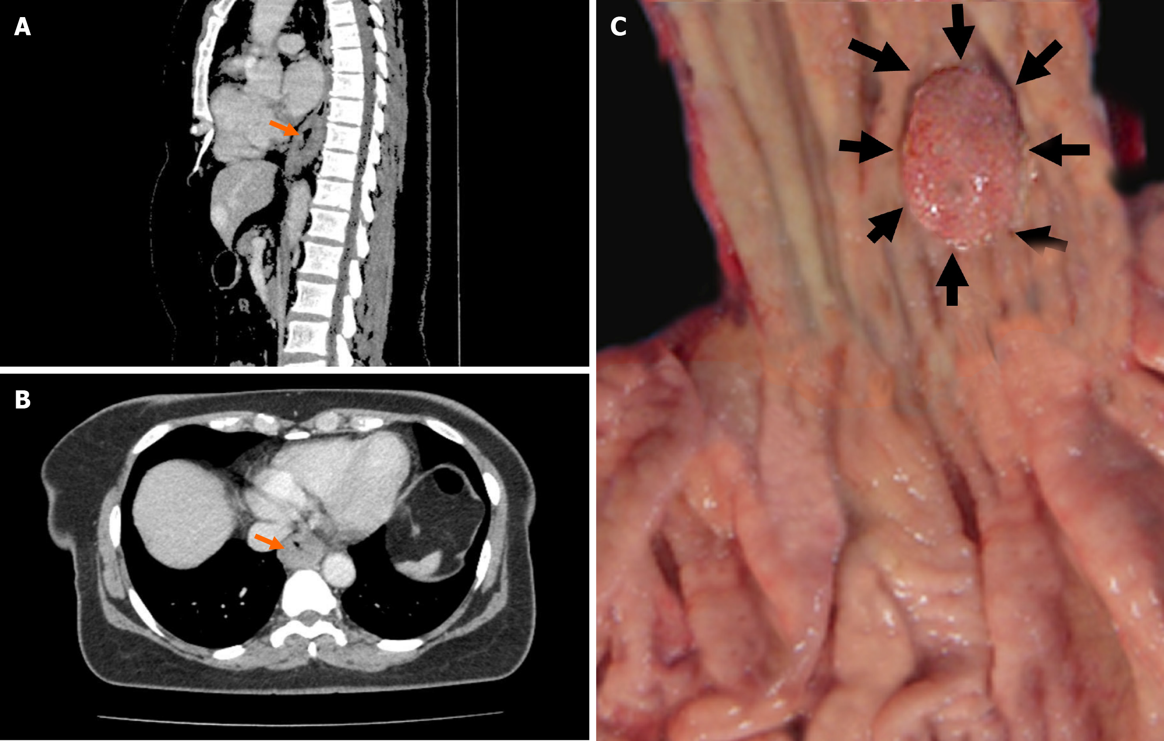
Figure 1 Contrast enhanced computed tomography and macroscopic findings.
A: In the contrast enhanced computed tomography, the sagittal section shows wall thickening in the distal esophagus (orange arrow); B: The axial section reveals wall thickening in the same localization. No lymph node metastasis is observed in either section; C: The macroscopic appearance of the lesion reveals its proximity to the gastroesophageal junction. This location is significant because it can influence both the surgical approach and the overall treatment strategy, given the anatomical and functional importance of the gastroesophageal junction.
- Citation: Yolburun SB, Gunizi OC, Elpek GO. Insights from literature of esophageal mucoepidermoid carcinoma in a female: A rare case report. World J Clin Cases 2025; 13(30): 110475
- URL: https://www.wjgnet.com/2307-8960/full/v13/i30/110475.htm
- DOI: https://dx.doi.org/10.12998/wjcc.v13.i30.110475