
















©The Author(s) 2025.
World J Clin Cases. Oct 26, 2025; 13(30): 110330
Published online Oct 26, 2025. doi: 10.12998/wjcc.v13.i30.110330
Published online Oct 26, 2025. doi: 10.12998/wjcc.v13.i30.110330
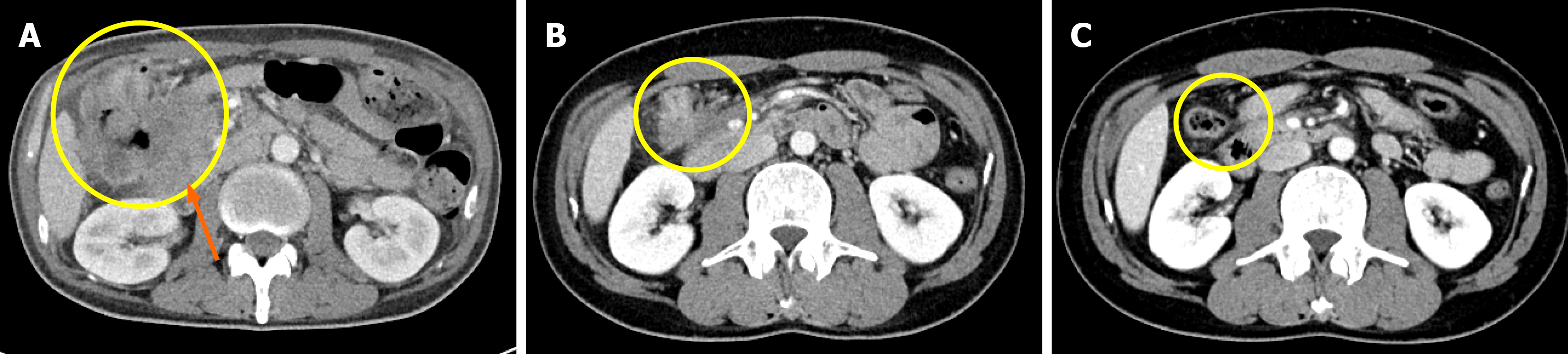
Figure 3 Axial Abdominopelvic computed tomography during the treatment course.
A: Baseline imaging revealed a large soft tissue mass in the proximal transverse colon with invasion into adjacent structures; B: After 6 months of pembrolizumab treatment, a marked reduction in tumor size was revealed; C: After 20 months of pembrolizumab treatment, radiologic complete response of the primary lesion was observed. The yellow circles indicate the location of the primary tumor across the treatment timeline. The orange arrow indicates the primary tumor in the colon with suspected invasion into the pancreas.
- Citation: Lee C, Kim MH, Choi ET, Park IJ, Lim SB, Yoon YS, Kim CW, Lee JL, Park EJ. Spontaneous colonic transection following pathologic complete response to pembrolizumab in high microsatellite instability colorectal cancer: A case report and review of literature. World J Clin Cases 2025; 13(30): 110330
- URL: https://www.wjgnet.com/2307-8960/full/v13/i30/110330.htm
- DOI: https://dx.doi.org/10.12998/wjcc.v13.i30.110330