
















©The Author(s) 2025.
World J Clin Cases. Oct 26, 2025; 13(30): 110314
Published online Oct 26, 2025. doi: 10.12998/wjcc.v13.i30.110314
Published online Oct 26, 2025. doi: 10.12998/wjcc.v13.i30.110314
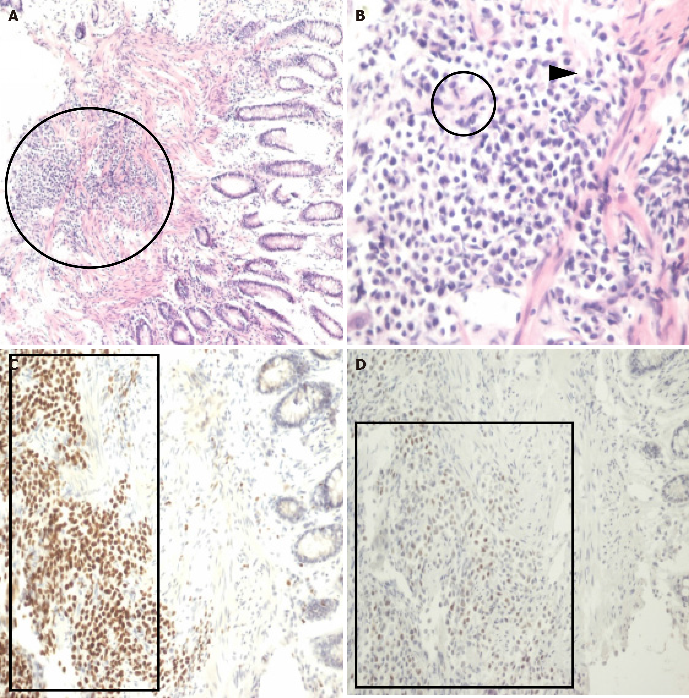
Figure 3 Histopathologic findings.
A: Discohesive tumor cells infiltrating the muscularis mucosa of the colon (black circle; hematoxylin–eosin staining, × 100); B: Plasmacytoid cells with abundant eosinophilic cytoplasm (black circle) and eccentric nuclei (black arrowhead; hematoxylin–eosin staining, × 400); C: Immunohistochemical staining for GATA-3 indicated nuclear positivity (black box; immunoperoxidase method, × 200); D: P63 staining revealed nuclear positivity (black box; immunoperoxidase method, × 200).
- Citation: Chen GY, Tsai HL, Chen PJ, Chen YC, Wang JY, Lin CY, Yin HL. Plasmacytoid urothelial carcinoma of the urinary bladder with colorectal metastasis and mimicking colitis: A case report. World J Clin Cases 2025; 13(30): 110314
- URL: https://www.wjgnet.com/2307-8960/full/v13/i30/110314.htm
- DOI: https://dx.doi.org/10.12998/wjcc.v13.i30.110314