
















©The Author(s) 2025.
World J Clin Cases. Oct 26, 2025; 13(30): 110314
Published online Oct 26, 2025. doi: 10.12998/wjcc.v13.i30.110314
Published online Oct 26, 2025. doi: 10.12998/wjcc.v13.i30.110314
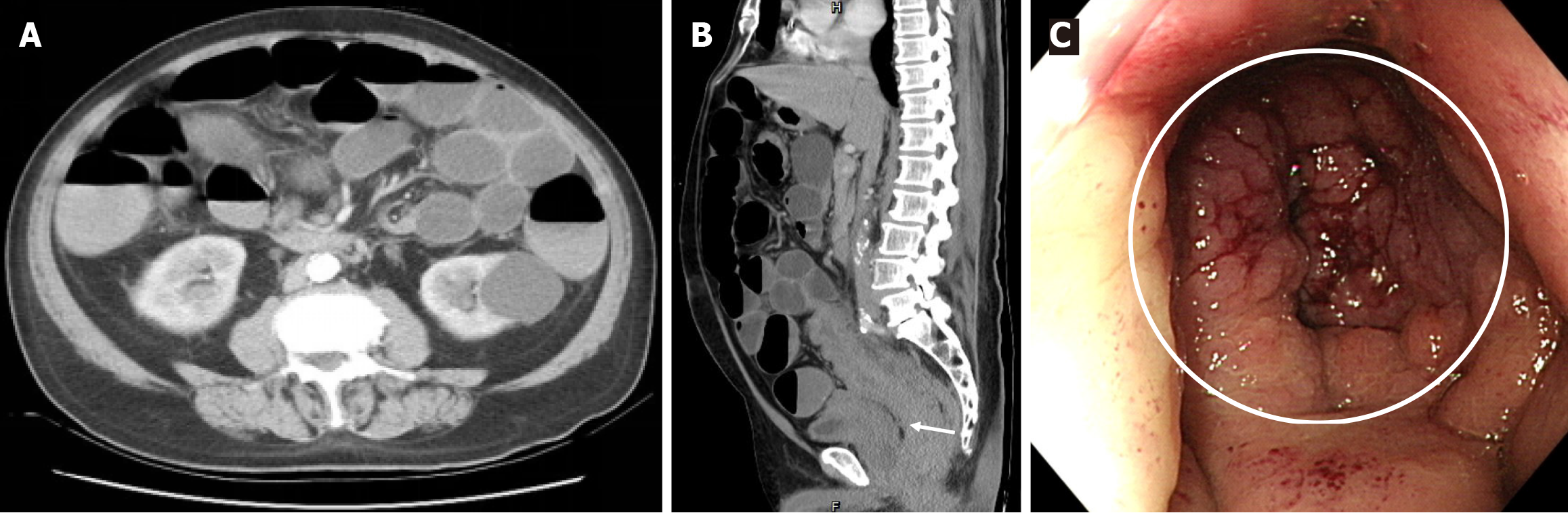
Figure 1 Preoperative evaluation.
A: Diffuse distension of the small and large intestines on axial-view abdominal computed tomography; B: Long segmental thickening of the rectosigmoid colonic wall and perirectal fat stranding with a clear vesicorectal fat plane (white arrow) on sagittal-view abdominal computed tomography; C: Obstructed lumen with mucosal swelling (white circle) located up to 5 cm from the anal verge on colonoscopy.
- Citation: Chen GY, Tsai HL, Chen PJ, Chen YC, Wang JY, Lin CY, Yin HL. Plasmacytoid urothelial carcinoma of the urinary bladder with colorectal metastasis and mimicking colitis: A case report. World J Clin Cases 2025; 13(30): 110314
- URL: https://www.wjgnet.com/2307-8960/full/v13/i30/110314.htm
- DOI: https://dx.doi.org/10.12998/wjcc.v13.i30.110314